-
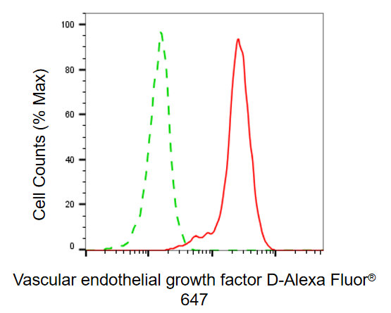
Flow cytometric analysis of vascular endothelial growth factor D expression in HepG2 cells using vascular endothelial growth factor D antibody. Green, isotype control; red, vascular endothelial growth factor D.
-
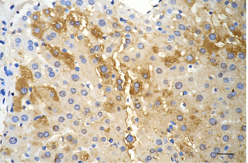
Immunohistochemistry was performed on paraffin-embedded mouse liver using vascular endothelial growth factor D antibody. Antigen retrieval was done in sodium citrate buffer (pH 6.0). DAB was used for detection, with hematoxylin counterstaining. Images were acquired using a Nikon Ci-L Plus microscope (40× objective). Scale bar: 25 μm.
-
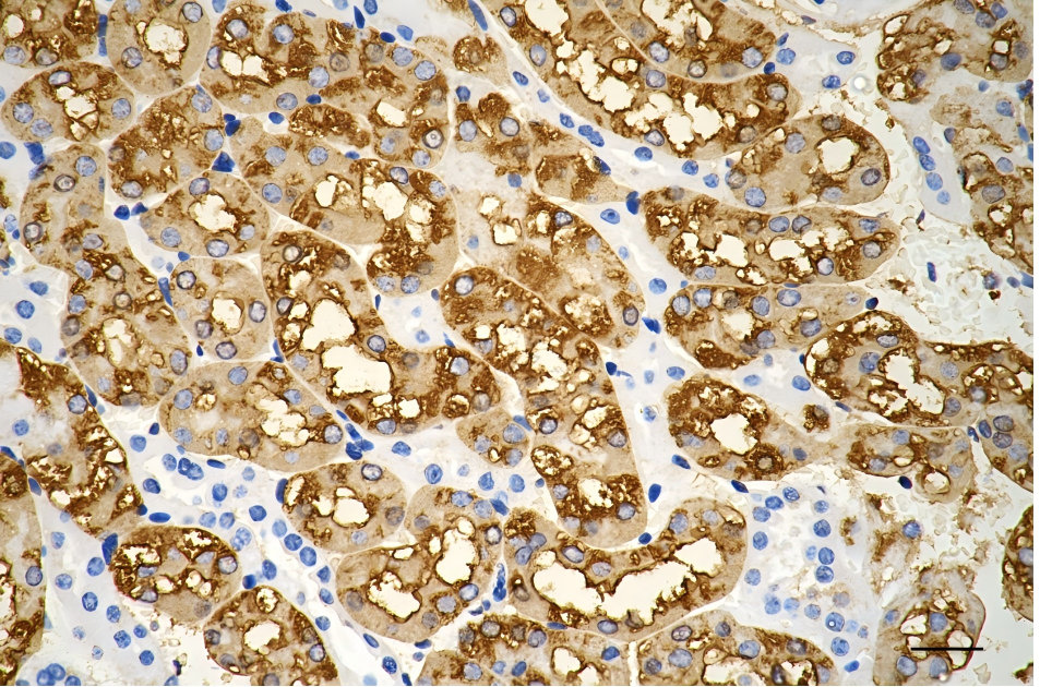
Immunohistochemistry was performed on paraffin-embedded mouse kidney using vascular endothelial growth factor D antibody. Antigen retrieval was done in sodium citrate buffer (pH 6.0). DAB was used for detection, with hematoxylin counterstaining. Images were acquired using a Nikon Ci-L Plus microscope (40× objective). Scale bar: 25 μm.
-
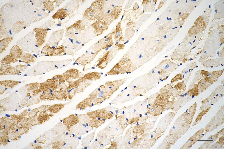
Immunohistochemistry was performed on paraffin-embedded mouse heart using vascular endothelial growth factor D antibody. Antigen retrieval was done in sodium citrate buffer (pH 6.0). DAB was used for detection, with hematoxylin counterstaining. Images were acquired using a Nikon Ci-L Plus microscope (40× objective). Scale bar: 25 μm.
-
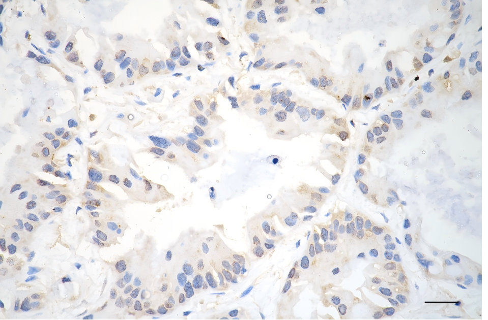
Immunohistochemistry was performed on paraffin-embedded human lung adenocarcinoma using vascular endothelial growth factor D antibody. Antigen retrieval was done in sodium citrate buffer (pH 6.0). DAB was used for detection, with hematoxylin counterstaining. Images were acquired using a Nikon Ci-L Plus microscope (40× objective). Scale bar: 25 μm.
-
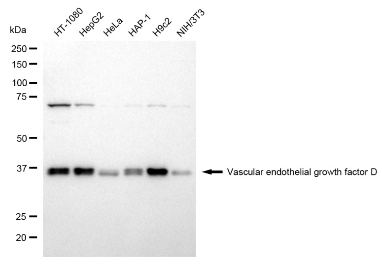
Western blotting analysis using vascular endothelial growth factor D antibody. Total cell lysates (30 μg) from various cell lines were loaded and separated by SDS-PAGE. The blot was incubated with vascular endothelial growth factor D antibody and HRP-conjugated goat anti-rabbit secondary antibody respectively.