Breast cancer is the leading cause of cancer death among women worldwide. The vast majority of breast cancers are carcinomas that originate from cells lining the milk-forming ducts of the mammary gland. Signs of breast cancer may include a lump in the breast, a change in breast shape, dimpling of the skin, fluid coming from the nipple, or a red scaly patch of skin. Risk factors for developing breast cancer include being female, obesity, lack of physical exercise, drinkingalcohol, hormone replacement therapy during menopause, ionizing radiation, early age at first menstruation, having children late or not at all, older age, and family history. About 5–10% of cases are due to genesinherited from a person's parents, including BRCA1 and BRCA2 among others.
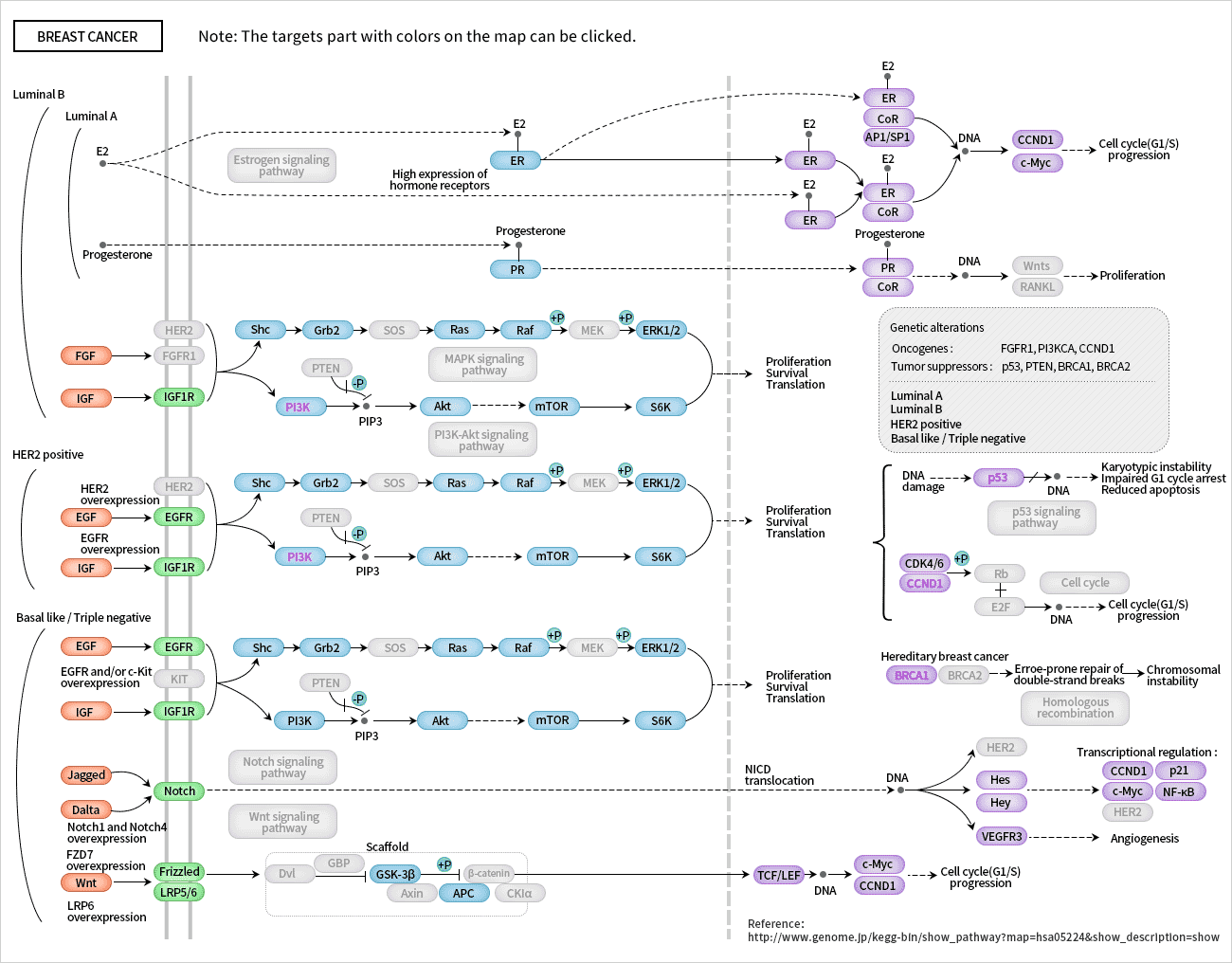
As the following figure shows, we summarize the breast cancer pathway and aim to help us in the research of breast cancer, including causes identification and the development of strategies for prevention, diagnosis, treatments and cure. Cusabio has a sound platform for the development of ELISA kit, mature antigen-antibody research and development system. Now we offer 178 ELISA kits in high specificity, high sensitivity, high stability and different species for your breast cancer research. Some of our products has cited by publications, such as HER2, HES1, WNT10B, and so on. We also have other products such as gene, protein and antibody for breast cancer research, For complete Elisa kits catalog, please visit https://www.cusabio.com/catalog-11-1.html.
If you are in need of other products such as gene, protein and antibody for breast cancer research, please feel free to contact us.
| Product Name | Code | Sample Type | Detect Range | Sensitivity |
|---|---|---|---|---|
| Human TP53 ELISA Kit | CSB-E08334h | serum, cell culture supernates, urine, cerebrospinal fluid (CSF), tissue homogenates, cell lysates | 9.38 pg/ml-600 pg/ml | 2.34 pg/ml |
| Rat TP53 ELISA Kit | CSB-E08336r | serum, plasma, tissue homogenates | 12.5 pg/ml-800 pg/ml | 3.12pg/ml |

Human epidermal growth factor receptor 2 (sp185/HER2) ELISA Kit(CSB-E11161h)purchased from cusabio.
VEGF, SDF-1α and HER2 serum levels were assessed by ELISA, both at baseline and during adjuvant treatment. At baseline, the VEGF and SDF-1α mean values were within the normal reference ranges, while the HER2 mean values showed an increase compared to the reference range. After AC-based therapy the VEGF level showed a significant increase (p = 0.011), while the SDF-1α and HER2 levels showed significant decreases (p = 0.001 and p = 0.004), respectively, compared to baseline values. At the end of the 12th cycle of taxane-based therapy, the VEGF level again exhibited a significant increase (p = 0.037), while the SDF-1α and HER2 levels were found to decrease again, although this decrease was significant only for SDF-1α (p= 0.003). The serum cytokine data, with their mean values (± SD) and pvalues, after AC-based and at the end of taxane-based therapy versus baseline, are summarized in Table 6. Additionally, we analyzed relationships between VEGF levels and the absolute numbers of CECs and their subsets and CEPs, but no direct correlations were detected.

Human NF-κB ELISA Kit (CSB-E12107h) and Human HES1 ELISA kit (CSB-EL010307HU) purchased from cusabio.
As shown in Figure 4, RANKL stimulation for 24 h obviously up-regulated the expressions of TGF-β1, NF-κB and Hes1 (P <0.01). Zoledronic acid and 0.08 mmol/L brucine markedly inhibited the expressions of TGF-β1, NF-κB and Hes1 compared with the RANKL-treated group (P<0.05 or P<0.01).
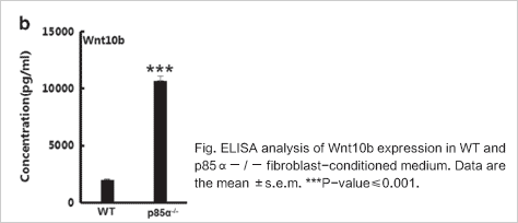
Human WNT10B ELISA Kit (CSB-EL026130HU) purchased from cusabio.
Wnt10b acts as a paracrine factor and has a crucial role in inducing breast cancer epithelial cell EMT and facilitating metastasis. Next, we measured the expression of Wnt family proteins involved in canonical pathways in p85α− / − fibroblasts and found that the expression of Wnt10b were significantly increased in p85α− / − fibroblasts compared with WT fibroblasts.
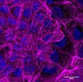
A study recently published in Nature Communications shows that breast cancer cells undergo a stiffening state prior to becoming malignant. The discovery, made by a research team led by Florence Janody, from Instituto Gulbenkian de Ciencia (IGC; Portugal), identifies a new signal in tumor cells that could be applicable in the design of cancer-targeting therapies.
Tavares, S., Vieira, A.F., Taubenberger, A.V. et al. Actin stress fiber organization promotes cell stiffening and proliferation of pre-invasive breast cancer cells. Nature Communications, Published online:16 May 2017, doi:10.1038/ncomms15237
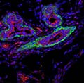
Researchers from Princeton University's Department of Molecular Biology have identified a small RNA molecule that helps maintain the activity of stem cells in both healthy and cancerous breast tissue. The study, which will be published in the June issue of Nature Cell Biology, suggests that this "microRNA" promotes particularly deadly forms of breast cancer and that inhibiting the effects of this molecule could improve the efficacy of existing breast cancer therapies.
Toni Celià-Terrassa et al, Normal and cancerous mammary stem cells evade interferon-induced constraint through the miR-199a–LCOR axis, Nature Cell Biology (2017). DOI: 10.1038/ncb3533
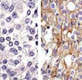
While target therapies directed toward genetic mutations that drive a tumor's growth have significantly improved the outlook for many patients, they have not been as successful in controlling brain metastases in several types of cancer. In the case of breast cancer driven by overexpression of the HER2 gene, up to 50 percent of patients treated with targeted therapies eventually develop brain metastases, which are inevitably fatal. Now a Massachusetts General Hospital (MGH)-based research team has identified a novel mechanism behind the resistance to HER2- or PI3K-targeted therapies and a treatment strategy that may overcome this resistance.
D.P. Kodack et al., The brain microenvironment mediates resistance in luminal breast cancer to PI3K inhibition through HER3 activation. Science Translational Medicine (2017).

A City of Hope-led study found that the use of low-dose aspirin (81mg) reduces the risk of breast cancer in women who are part of the California's Teacher's Study. This study—which is the first to suggest that the reduction in risk occurs for low-dose aspirin—was proposed by City of Hope's Leslie Bernstein, Ph.D., professor and director of the Division of Biomarkers of Early Detection and Prevention, and published online in the journal, Breast Cancer Research.
Christina A. Clarke et al, Regular and low-dose aspirin, other non-steroidal anti-inflammatory medications and prospective risk of HER2-defined breast cancer: the California Teachers Study,Breast. Cancer Research (2017). DOI: 10.1186/s13058-017-0840-7