Call us
301-363-4651 (Available 9 a.m. to 5 p.m. CST from Monday to Friday)
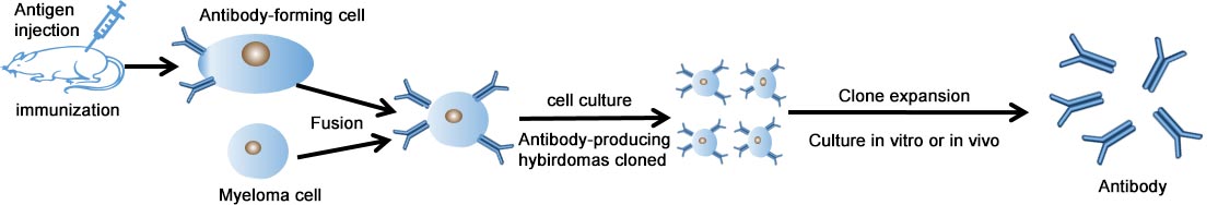
Cusabio monoclonal antibodies are made by identical immune cells from a unique parent cell, which have better affinity compared with the same kind polyclonal antibodies.
Cusabio could offer a reliable production of custom monoclonal antibodies upon customers’ various demands. Based on large-scale production as well as high quality, Cusabio custom monoclonal antibodies have been widely used in screening therapeutic targets and drugs discovery.
The production process of Cusabio custom monoclonal antibody is as follows.
Process & Deliverables:
| Immunogen Options | Process | Deliverables | Production Time |
|---|---|---|---|
| Peptide |
|
|
18-22 Weeks |
| Recombinant Protein | 20-24 Weeks | ||
| Native Protein | 20-24 Weeks | ||
| mRNA-LNP | 20-24 Weeks |
Successful Showcase (partial)
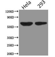
Western Blot
Positive WB detected in:Hela whole cell lysate,HEK293 whole cell lysate
All lanes : PODXL antibody at 2.5 ug/ml
Secondary
Goat polyclonal to Mouse IgG at 1/5000 dilution
Predicted band size: 59KDa, 56KDa
Observed band size: 59KDa
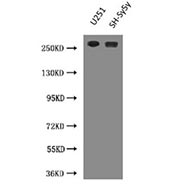
Western Blot
Positive WB detected in:U251 whole cell lysate,SH-Sy5y whole cell lysate
All lanes :NES antibody at 3 ug/ml
Predicted band size: 260KDa
Observed band size: 260KDa
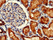
IHC image of A antibody diluted at 1:100 and staining in paraffin-embedded human kidney tissue performed on a Leica BondTM system. After dewaxing and hydration, antigen retrieval was mediated by high pressure in a citrate buffer (pH 6.0). Section was blocked with 10% normal goat serum 30min at RT. Then primary antibody (1% BSA) was incubated at 4°C overnight. The primary is detected by a biotinylated secondary antibody and visualized using an HRP conjugated SP system.
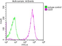
Overlay histogram showing THP-1 cells stained with CD31 (red line) at 1:500. The cells were incubated in 1x PBS /10% normal goat serum to block non-specific protein-protein interactions followed by primary antibody for 1 h at 4℃.The secondary antibody used was FITC goat anti-mouse IgG (H+L) at 1/200 dilution for 1 h at 4℃. Isotype control antibody (green line) was used under the same conditions. Acquisition of >10,000 events was performed.
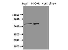
Immunoprecipitating PODXL in HEK293 whole cell lysate
Lane 1: Rabbit monoclonal IgG(1 ug)instead of PODXL in HEK293 whole cell lysate. For western blotting,a HRP-conjugated Protein G antibody was used as the secondary antibody (1/2000)
Lane 2: PODXL(8 ug)+ HEK293 whole cell lysate(500 ug)
Lane 3: HEK293 whole cell lysate (10 ug)
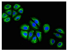
Immunofluorescence staining of A549 cells with A antibody at 1:100, counter-stained with DAPI. The cells were blocked in 10% normal Goat Serum and then incubated with the primary antibody overnight at 4°C.The secondary antibody was Alexa Fluor 488-congugated AffiniPure Goat Anti-Mouse IgG (H+L).
Send an Inquiry
You can contact us by email, telephone or online
Specialized CRO Services

