Call us
301-363-4651 (Available 9 a.m. to 5 p.m. CST from Monday to Friday)
CUSABIO offers a reliable and wide range of custom polyclonal antibody production with multiple immunogen options as well as multiple host species options.
The production process of Cusabio custom polyclonal antibody is as follows.
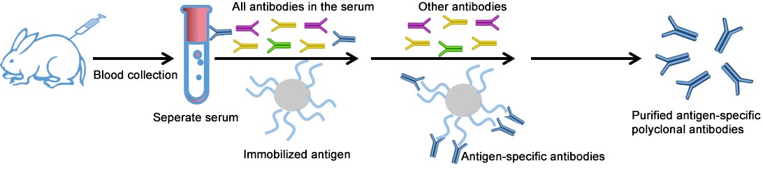
Process & Deliverables:
| Immunogen Options | Process | Deliverables | Production Time |
|---|---|---|---|
| Peptide |
|
|
12-14 Weeks |
| Recombinant Protein | 14-16 Weeks | ||
| Native Protein | 12-14 Weeks | ||
| mRNA-LNP | 14-16 Weeks |
Successful Showcase (partial)
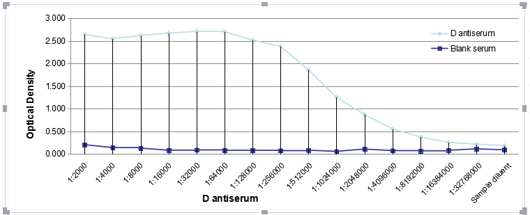
ELISA
Antigen coating concentration 2 ug/ml
Antiserum 1:2000 is more than diluted
Secondary:Goat polyclonal to rabbit IgG at 1/50000 dilution
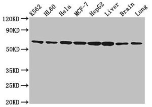
Western Blot
Positive WB detected in:K562 whole cell lysate,HL-60 whole cell lysate,Hela whole cell lysate, MCF-7 whole cell lysate, HepG2 whole cell lysate,Rat liver tissue, Mouse brain tissue, Mouse lung tissue
All lanes:a antibody at 2ug/ ml
Secondary
Goat polyclonal to rabbit IgG at 1/50000 dilution
Predicted band size: 64 KDa
Observed band size: 64 KDa
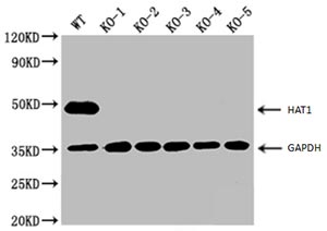
Western blot
WT: Wild-type 293 cells
KO: Knockout 293 cells
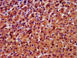
IHC image of A antibody diluted at 1:400 and staining in paraffin-embedded human adrenal gland tissue performed on a Leica BondTM system. After dewaxing and hydration, antigen retrieval was mediated by high pressure in a citrate buffer (pH 6.0). Section was blocked with 10% normal goat serum 30min at RT. Then primary antibody (1% BSA) was incubated at 4°C overnight. The primary is detected by a biotinylated secondary antibody and visualized using an HRP conjugated SP system.
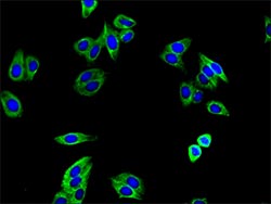
Immunofluorescence staining of HepG2 cells with A antibody at 1:400,counter-stained with DAPI. The cells were fixed in 4% formaldehyde, permeabilized using 0.2% Triton X-100 and blocked in 10% normal Goat Serum. The cells were then incubated with the antibody overnight at 4°C.The secondary antibody was Alexa Fluor 488-congugated AffiniPure Goat Anti-Rabbit IgG (H+L).
Send an Inquiry
You can contact us by email, telephone or online
Specialized CRO Services

