Call us
301-363-4651 (Available 9 a.m. to 5 p.m. CST from Monday to Friday)
Free samples available for a limited time.
Request a Free Sample NowThe efficiency of antibody internalization is key to designing effective antibody-drug conjugates (ADCs). For the cytotoxic drug to reach and destroy tumor cells, the antibody must first be internalized by the target cell. Without this step, the drug cannot be released where it's needed.
DT3C protein is a reliable tool for measuring how well antibodies are internalized. It helps researchers evaluate the internalization efficiency of antibodies in target cells, making it easier to identify those with strong binding and efficient internalization. These high-performing antibodies can be used in ADCs to improve drug targeting and safety.
To see why DT3C works, we need to explore its structural and functional features.
DT3C protein is a recombinant fusion protein that consists of a diphtheria toxin (DT) protein lacking the receptor-binding domain (only catalytic and translocation domains) but containing the C1, C2, and C3 domains (IgG-binding domain) of Streptococcus protein G (3C). It is characterized by low toxicity and high affinity with antibodies.
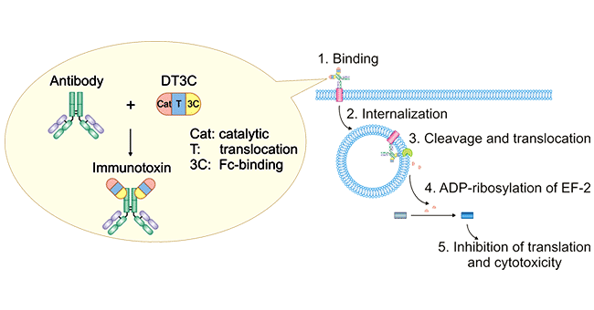
Figure 1. Mechanism of antibody: DT3C-induced cytotoxicity
DT3C's unique structure enables its role in mAb-DT3C conjugates, which mimic ADC behavior in vitro.
A mAb-DT3C conjugate, which functions in vitro similarly to an antibody-drug conjugate (ADC), can kill cancer cells or reduce the viability of cancer cells only when the mAb being tested is internalized by the target cells through antibody-antigen interactions.
After the mAb-DT3C conjugate recognizes and binds to cell surface antigens, the mAb-DT3C-antigen complex is internalized into the cell via endocytosis. In the cell, the translocated terminus of DT3C is cleaved by the cellular furin protease, and the catalytic domain of DT3C is released into the cytoplasm, leading to ADP ribosylation of EF-2, subsequent blockade of the protein translation machinery, and ultimate cytotoxicity.
Therefore, the internalization efficiency of antibodies can be evaluated according to the cell-killing situation.
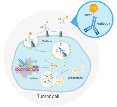
Figure 2. DT3C-Induced Cell Killing Mechanism
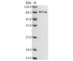
CUSABIO DT3C recombinant protein (CSB-EP360556CQR1) was expressed from E.coli, containing 3 fragments (33-417aa & a linker & 291-497aa) and carrying the 6*His tag sequence placed on the N-terminal of the DT3C coding sequence.
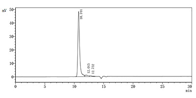
The purity of the recombinant DT3C protein was assessed using SDS-PAGE and SEC-HPLC, demonstrating a purity level of up to 90%. DT3C migrated as a band on the gel with an approximate molecular weight of 69.4 kDa. Dissolved in PBS with pH7.4 and sterilized by 0.2μm filtration membrane, this DT3C’s endotoxin is < 1.0 EU/ug as determined by the LAL method.
After incubating different concentrations of DT3C protein with CCR8 antibody at 37°C for 30 minutes, the mixture was added to a culture plate and incubated for 48 hours. The range of EC50 values are 0.5795-1.400 μg/mL and 0.5608-1.318 μg/mL, respectively.
In vitro evaluation of antibody internalization efficiency
ADC drug research
Antibody screening and evaluation
Simple and Efficient Operation: mAb-DT3C conjugates can be formed by incubating at room temperature for 30 minutes. These conjugates function similarly to actual antibody-drug conjugates (ADCs) in vitro, making it effective for screening monoclonal antibodies capable of cellular internalization.
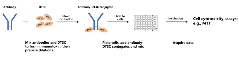
Antibody Internalization Assay Workflow
Multi-species Applicability: DT3C can bind to IgG from various species, including human, mouse, rabbit, goat, etc., to form mAb-DT3C conjugates. This versatility makes it suitable for experiments involving antibodies from different sources.
High Precision: mAb-DT3C conjugates significantly reduce cell viability only when internalized by target cells. This ensures DT3C accurately identifies antibodies that can truly undergo cellular internalization.
High internalization efficiency: Compared to Mab-ZAP, DT3C offers higher efficiency and precision in screening internalized antibodies. Mab-ZAP's larger molecular size can interfere with antibody internalization, an issue that DT3C effectively avoids.
In vitro detection of ADC antibody internalization efficiency
Reference: M. Yamaguchi et al. Biochemical and Biophysical Research Communications 454 (2014) 600-603.
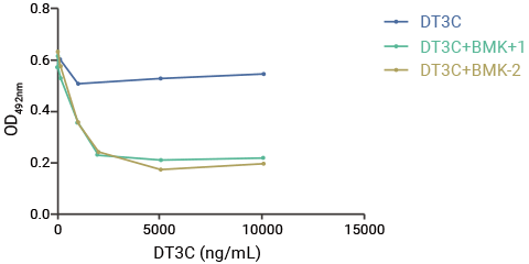
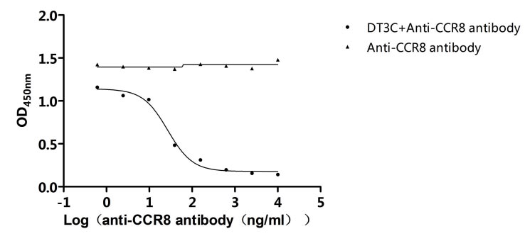
Request a Free Sample NowInternalization of anti-CCR8-DT3C
Method:
Different concentrations of anti-CCR8 antibody (CSB-RA004847MA3HU) were incubated with DT3C protein his tag (CSB-EP360556CQR1) at 37℃ for 30min, and then added to the the 96-well microplates with CHO-K1/Human CCR8 Stable Cell Line cell suspension of the same volume, respectively, for 48h at 37℃. Cell viability was detected using a CCK-8 kit.
Result:
The ED50 value is 15.92-45.59 ng/ml. As shown in the Cytotoxicity Assay (the following figure), anti-CCR8 mAb and DT3C were successfully internalized and transported to the lysosomes. This indicates that the DT3C protein demonstrates high antibody internalization efficiency.
Generally speaking, antibody internalization assays primarily include pH probe-based assays, live-cell imaging-based assays, temperature shift-based assays, and toxin-conjugated cytotoxicity assays.
Among these methods, temperature shift-based assays have room for improvement in accuracy and specificity. Live-cell imaging-based assay, on the other hand, require advanced equipment, which limits its accessibility. Toxin-conjugated cytotoxicity assays have had relatively few well-established products in the past.
As a result, pH probe-based assays remain the most commonly used method for antibody internalization today.
pH probe-based assays may be affected by other intracellular factors that influence fluorescent signals. However, they are affordable, simple, and efficient. These advantages make them highly favored by researchers. As a result, they often overshadow newer methods for antibody internalization detection—for example, the DT3C protein, as mentioned earlier.
Similar to the well-known mAb-ZAP, DT3C is also a toxin-conjugated cytotoxicity assay. It matches the pH probe in performance for naked antibody screening and offers a key advantage—it can be used during the early-stage supernatant screening of hybridomas, significantly accelerating the initial stages of ADC development. This is something neither pH probe-based methods nor mAb-ZAP can achieve.
Compared to mAb-ZAP, DT3C protein offers more distinct advantages for antibody internalization assays. Research by M. Yamaguchi et al. has shown that DT3C performs significantly better than mAb-ZAP in antibody internalization efficiency. Let’s take a closer look at the comparison between these two toxin-conjugated cytotoxicity methods.
| Methods | Detection Methods | Equipments Required | Applicable Scenarios | Features | Versatility | Duration of Experiment | Starting prices |
|---|---|---|---|---|---|---|---|
| mAb-ZAP | Cytotoxicity Assay | ELISA Reader |
|
|
Low | 4-5 days | $550/ 25µg |
| DT3C protein | Cytotoxicity Assay | ELISA Reader |
|
|
High | 2-3 days | $388/ 20µg |
DT3C binds to Fc terminal of the antibody.
Cell viability is measured using MTT or CTG assays, and CCK8 is also an option.
It can take 24, 48, or 72 hours, depending on the cell proliferation rate.
66 kDa.
This has not been tested yet. Theoretically, conjugating DT3C does not affect the antibody's stability.
DT3C binds to the Fc region of antibodies via the 3C domain of Streptococcus protein G, similar to protein G. Serum contains IgG, which can bind DT3C and interfere with the results, so serum-free medium is required for certain steps.
Lyophilized from 0.2 μm sterile-filtered PBS, 6% trehalose, pH 7.4.
Theoretically, concentration is not affected, but there might be a slight loss in yield during the process.
DT3C specifically binds to the Fc region of antibodies via protein G. DT3C alone is non-toxic to cells.
DT3C requires an Fc region to bind. If your nanobody has an Fc tag, it can work; otherwise, it is not applicable.
DT3C is mainly used to bind candidate antibodies, simulating ADC antibodies, to test their internalization efficiency. However, ADC antibodies inherently induce internalization, so their efficiency can be evaluated without DT3C.
Experimental steps: