SARS-CoV-2 S1 RBD ELISA Kit

Code CSB-EL33244
Size 96T,5×96T,10×96T
Price Request a Quote or Start an on-line Chat
Trial Size 24T ELISA Kit Trial Size (Only USD$150/ kit)
* The sample kit cost can be deducted from your subsequent orders of 96T full size kits of the same analyte at 1/5 per kit, until depleted in 6 months. Apply now

Product Details

Target Name
Novel Coronavirus Spike Glycoprotein RBD (SARS-CoV-2 S1 RBD)
Alternative Names
S; 2; Spike glycoprotein; S glycoprotein; E2; Peplomer protein)
Abbreviation
SARS-CoV-2 S1 RBD
Uniprot No.
Sample Types
serum, plasma, swabs
Detection Range
1.25 ng/mL-80 ng/mL
Sensitivity
0.31 ng/mL
Assay Time
1-5h
Sample Volume
50-100ul
Detection Wavelength
450 nm
Research Area
Infectious Diseases
Assay Principle
quantitative
Measurement
Sandwich
Precision
Intra-assay Precision (Precision within an assay): CV%<8%  
Three samples of known concentration were tested twenty times on one plate to assess.
Inter-assay Precision (Precision between assays): CV%<10%  
Three samples of known concentration were tested in twenty assays to assess.

  Intra-Assay Precision Inter-Assay Precision
Sample 1 2 3 1 2 3
n 20 20 20 20 20 20
Mean(ng/ml) 10.79 11.06 11.36 10.85 10.99 10.02
SD 0.032 0.034 0.039 0.041 0.045 0.041
CV(%) 5.53 5.74 6.43 7.05 7.64 7.55
Typical Data

These standard curves are provided for demonstration only. A standard curve should be generated for each set of samples assayed.
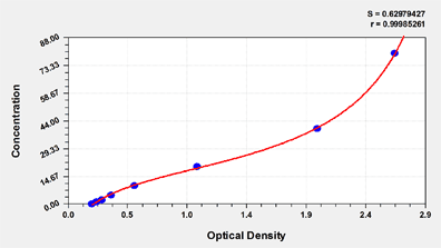
 

ng/ml OD1 OD2 Average Corrected
80 2.648 2.594 2.621 2.409
40 1.973 2.032 2.003 1.791
20 1.027 1.069 1.048 0.836
10 0.559 0.537 0.548 0.336
5 0.365 0.358 0.362 0.150
2.5 0.291 0.285 0.288 0.076
1.25 0.251 0.240 0.246 0.034
0 0.213 0.211 0.212


Troubleshooting
and FAQs
Storage
Store at 2-8°C. Please refer to protocol.
Lead Time
3-5 working days after you place the order, and it takes another 3-5 days for delivery via DHL or FedEx

Customer Reviews and Q&A

 Customer Reviews

There are currently no reviews for this product.

Submit a Review here

Target Background

Function
(From Uniprot)
attaches the virion to the cell membrane by interacting with host receptor, initiating the infection. Binding to human ACE2 receptor and internalization of the virus into the endosomes of the host cell induces conformational changes in the Spike glycoprotein. Binding to host NRP1 and NRP2 via C-terminal polybasic sequence enhances virion entry into host cell. This interaction may explain virus tropism of human olfactory epithelium cells, which express high level of NRP1 and NRP2 but low level of ACE2. The stalk domain of S contains three hinges, giving the head unexpected orientational freedom. Uses human TMPRSS2 for priming in human lung cells which is an essential step for viral entry. Can be alternatively processed by host furin. Proteolysis by cathepsin CTSL may unmask the fusion peptide of S2 and activate membranes fusion within endosomes.; mediates fusion of the virion and cellular membranes by acting as a class I viral fusion protein. Under the current model, the protein has at least three conformational states: pre-fusion native state, pre-hairpin intermediate state, and post-fusion hairpin state. During viral and target cell membrane fusion, the coiled coil regions (heptad repeats) assume a trimer-of-hairpins structure, positioning the fusion peptide in close proximity to the C-terminal region of the ectodomain. The formation of this structure appears to drive apposition and subsequent fusion of viral and target cell membranes.; Acts as a viral fusion peptide which is unmasked following S2 cleavage occurring upon virus endocytosis.; May down-regulate host tetherin (BST2) by lysosomal degradation, thereby counteracting its antiviral activity.
Gene References into Functions
  1. Study presents crystal structure of C-terminal domain of SARS-CoV-2 (SARS-CoV-2-CTD) spike S protein in complex with human ACE2 (hACE2); hACE2-binding mode similar overall to that observed for SARS-CoV. However, details at the binding interface show that key residue substitutions in SARS-CoV-2-CTD slightly strengthen the interaction and lead to higher affinity for receptor binding than SARS-CoV receptor-binding domain. PMID: 32378705
  2. crystal structure of the receptor-binding domain (RBD) of the spike protein of SARS-CoV-2 bound to the cell receptor ACE2 PMID: 32365751
  3. crystal structure of the receptor-binding domain (RBD) of the spike protein of SARS-CoV-2 (engineered to facilitate crystallization) in complex with ACE2 PMID: 32320687
  4. Out of the two isolates from India compared to the isolates from Wuhan, China, one was found to harbor a mutation in its receptor-binding domain (RBD) at position 407 where, arginine was replaced by isoleucine. This mutation has been seen to change the secondary structure of the protein at that region and this can potentially alter receptor binding of the virus. PMID: 32275855
  5. Structural modeling of the SARS-CoV-2 spike glycoprotein show similar receptor utilization between SARS-CoV-2 and SARS-CoV, despite a relatively low amino acid similarity in the receptor binding module. Compared to SARS-CoV and all other coronaviruses in Betacoronavirus lineage B, an extended structural loop containing basic amino acids were identified at the interface of the receptor binding (S1) and fusion (S2) domains. PMID: 32245784
  6. crystal structure of CR3022, a neutralizing antibody from a SARS patient, in complex with the receptor-binding domain of the SARS-CoV-2 spike (S) protein to 3.1 A; study provides insight into how SARS-CoV-2 can be targeted by the humoral immune response and revealed a conserved, but cryptic epitope shared between SARS-CoV-2 and SARS-CoV PMID: 32225176
  7. SARS-CoV and SARS-CoV-2 spike proteins have comparable binding affinities achieved by balancing energetics and dynamics. The SARS-CoV-2-ACE2 complex contains a higher number of contacts, a larger interface area, and decreased interface residue fluctuations relative to the SARS-CoV-ACE2 complex. PMID: 32225175
  8. Interaction interface between cat/dog/pangolin/Chinese hamster ACE2 and SARS-CoV/SARS-CoV-2 S protein was simulated through homology modeling. Authors identified that N82 of ACE2 showed closer contact with receptor-binding domain of S protein than human ACE2. PMID: 32221306
  9. SARS-CoV-2 S glycoprotein harbors a furin cleavage site at the boundary between the S1/S2 subunits, which is processed during biogenesis and sets this virus apart from SARS-CoV and SARS-related CoVs; determined cryo-EM structures of the SARS-CoV-2 S ectodomain trimer. PMID: 32201080
  10. Study demonstrates that SARS-CoV-2 uses the SARS-CoV receptor ACE2 for entry and the serine protease TMPRSS2 for S protein priming. PMID: 32155444
  11. The ACE2-B0AT1 complex exists as a dimer of heterodimers. Structural alignment of the RBD-ACE2-B0AT1 ternary complex with the S protein of SARS-CoV-2 suggests that two S protein trimers can simultaneously bind to an ACE2 homodimer. PMID: 32142651
  12. study demonstrated SARS-CoV-2 S protein entry on 293/hACE2 cells is mainly mediated through endocytosis, and PIKfyve, TPC2 and cathepsin L are critical for virus entry; found that SARS-CoV-2 S protein could trigger syncytia in 293/hACE2 cells independent of exogenous protease; there was limited cross-neutralization activity between convalescent sera from SARS and COVID-19 patients PMID: 32132184
  13. study determined a 3.5-angstrom-resolution cryo-electron microscopy structure of the 2019-nCoV S trimer in the prefusion conformation; provided biophysical and structural evidence that the 2019-nCoV S protein binds angiotensin-converting enzyme 2 (ACE2) with higher affinity than does severe acute respiratory syndrome (SARS)-CoV S PMID: 32075877

Show More

Hide All

Subcellular Location
Virion membrane; Single-pass type I membrane protein. Host endoplasmic reticulum-Golgi intermediate compartment membrane; Single-pass type I membrane protein. Host cell membrane; Single-pass type I membrane protein.
Protein Families
Betacoronaviruses spike protein family
icon of phone
Call us
301-363-4651 (Available 9 a.m. to 5 p.m. CST from Monday to Friday)
icon of address
Address
7505 Fannin St., Ste 610, Room 7 (CUBIO Innovation Center), Houston, TX 77054, USA
icon of social media
Join us with

Subscribe newsletter

Leave a message

* To protect against spam, please pass the CAPTCHA test below.
CAPTCHA verification
© 2007-2024 CUSABIO TECHNOLOGY LLC All rights reserved. 鄂ICP备15011166号-1
webinars: DT3C facilitates antibody internalization X