| Image |
-
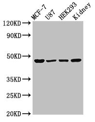
Western Blot
Positive WB detected in: MCF-7 whole cell lysate, U87 whole cell lysate, HEK293 whole cell lysate, Rat kidney tissue
All lanes: WWOX antibody at 3µg/ml
Secondary
Goat polyclonal to rabbit IgG at 1/50000 dilution
Predicted band size: 47, 42, 22, 5, 27, 36, 24 kDa
Observed band size: 47 kDa
-
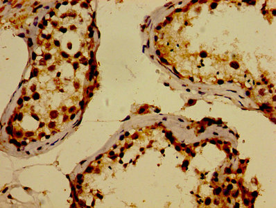
IHC image of CSB-PA873704LA01HU diluted at 1:300 and staining in paraffin-embedded human testis tissue performed on a Leica BondTM system. After dewaxing and hydration, antigen retrieval was mediated by high pressure in a citrate buffer (pH 6.0). Section was blocked with 10% normal goat serum 30min at RT. Then primary antibody (1% BSA) was incubated at 4°C overnight. The primary is detected by a biotinylated secondary antibody and visualized using an HRP conjugated SP system.
-
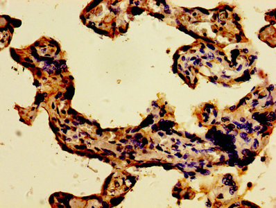
IHC image of CSB-PA873704LA01HU diluted at 1:300 and staining in paraffin-embedded human placenta tissue performed on a Leica BondTM system. After dewaxing and hydration, antigen retrieval was mediated by high pressure in a citrate buffer (pH 6.0). Section was blocked with 10% normal goat serum 30min at RT. Then primary antibody (1% BSA) was incubated at 4°C overnight. The primary is detected by a biotinylated secondary antibody and visualized using an HRP conjugated SP system.
-
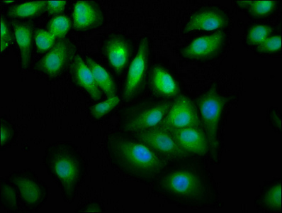
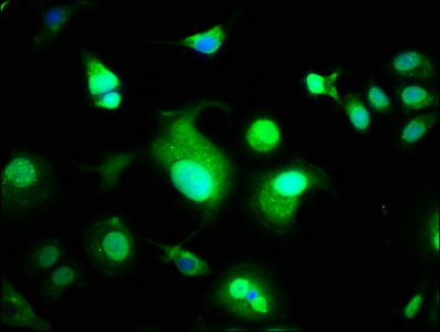
Immunofluorescence staining of A549 cells with CSB-PA873704LA01HU at 1:100, counter-stained with DAPI. The cells were fixed in 4% formaldehyde, permeabilized using 0.2% Triton X-100 and blocked in 10% normal Goat Serum. The cells were then incubated with the antibody overnight at 4°C. The secondary antibody was Alexa Fluor 488-congugated AffiniPure Goat Anti-Rabbit IgG(H+L).
-
Immunofluorescence staining of MCF-7 cells with CSB-PA873704LA01HU at 1:100, counter-stained with DAPI. The cells were fixed in 4% formaldehyde, permeabilized using 0.2% Triton X-100 and blocked in 10% normal Goat Serum. The cells were then incubated with the antibody overnight at 4°C. The secondary antibody was Alexa Fluor 488-congugated AffiniPure Goat Anti-Rabbit IgG(H+L).
-
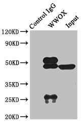
Immunoprecipitating WWOX in Rat kidney tissue
Lane 1: Rabbit control IgG instead of CSB-PA873704LA01HU in Rat kidney tissue. For western blotting, a HRP-conjugated light chain specific antibody was used as the secondary antibody (1/50000)
Lane 2: CSB-PA873704LA01HU (8µg) + Rat kidney tissue (500µg)
Lane 3: Rat kidney tissue (10µg)
|