Call us
301-363-4651 (Available 9 a.m. to 5 p.m. CST from Monday to Friday)
2. Pathways
2.1 Endogenous mitochondrial pathway
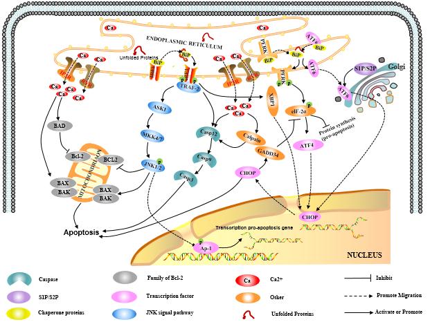
2.2 Endogenous endoplasmic reticulum pathway
Endoplasmic reticulum(ER) is the main site of protein processing, as well a major storage compartment of Ca2+ in cell, therefore, being decisively important in the synthesis, folding, modification and transport of protein, and maintaining Ca2+ homeostasis. The accumulation of unfold or misfolded proteins and disturbance of Ca2+ homeostasis in endoplasmic reticulum trigger the ER stress response (ERSR), which facilitates protein folding and removal of damaged proteins, and stabilizes ER Ca2+ homeostasis, but excessive ER stress response triggers apoptotic signals and induces apoptosis.
2.2.1 Regulation of apoptosis by the misfolded protein response
In eukaryotic cells, the unfolded protein response (UPR) is a self-protective mechanisms that responds to ER stress. The highly intense and prolonged UPR triggers three transmembrane protein PERK, IREI and ATF6 to repair cells, and samutalneously induces three ER stress-mediated apoptosis pathways.
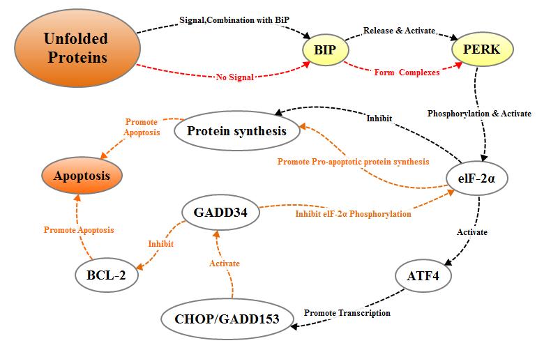
2.2.1.1 PERK pathway
PERK is a transmembrane protein kinase of the PEK family resident in the endoplasmic reticulum (ER) membran. When proteins are properly folded, PERK is in combination with formolecular chaperone like BiP/GRP78 to form stable compound, the combination of misfolded proteins and BiP/GRP78 will interfere with the interaction between PERK and BiP/GRP78. The released PERK is activated by oligomerization and reverse autophosphorylation, and activated PERK phosphorylates the alpha subunit of translation initiation factor 2 (eIF-2a). In the early stage of stress response, phosphorylated eIF2α inhibits the translation and synthesis of proteins and reduces the load of protein folding in the endoplasmic reticulum, thereby protecting the cells. With the increase of the intensity and duration of stress reaction, phosphorylated eIF-2α induces the transcriptional expression of transcription factor ATF4, and ATF4 can promote the expression of apoptotic signal molecule CHOP / GADD153, which in turn tiggers cell apoptosis.
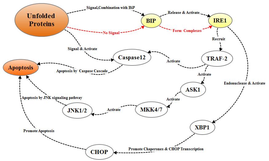
2.2.1.2 IREI pathway
IREI is another protein kinase located on the endoplasmic reticulum membrane. The IREI signal pathway is activated in the same way as PERK. When unfolded proteins accumulate in the endoplasmic reticulum, the IREI-BIP / GRP78 complex dissociates, the released IREI becomes oligomerized and activated after reverse autophosphorylation; The activated IREI can transmit cell survival signal and apoptosis signal. During the process of apoptosis, the activated IRE1 recruits cytosolic regulatory protein TRAF-2, indirectly recruiting and activating c-Jun N-terminal kinase, which inhibits apoptosis inhibitor proteins of the Bcl-2 family through phosphorylation. On the other hand, activated TRAF-2 simultaneously activates Caspase12 and initiates caspase cascade to mediate apoptosis. In addition, IRE1 also has ribonuclease activity which cleaves XBP1 mRNA to promotes the maturation of XBP1 mRNA and enhances the transcriptional expression of molecular chaperone protein and CHOP / GADD153, thereby promoting apoptosis.
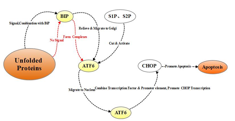
2.2.1.3 ATF6 pathway
ATF6 is a type II transmembrane protein located on the endoplasmic reticulum. The N-terminal intracellular domain of ATF6 contains the DNA transcriptional activation domain and nuclear localization signal of b-ZIP. Under non-stress conditions, ATF6 resides in the endoplasmic reticulum membrane in the form of zymogen, and under endoplasmic reticulum stress, ATF6 is transported to the Golgi apparatus via vesicles, where it undergoes cleavage by site-1 and site-2 (S1P and S2P) proteases and then relocate to the nucleus with the nuclear localization signal, inducing transcriptional expression of the endoplasmic reticulum stress gene including CHOP / GADD153 in the nucleus.
Both PERK, IRE1 and ATF6 signal pathways can induce the expression of CHOP/GADD153, as a direct result of of endoplasmic reticulum stress. CHOP/GADD153 plays an important role in cell growth arrest and cell death.
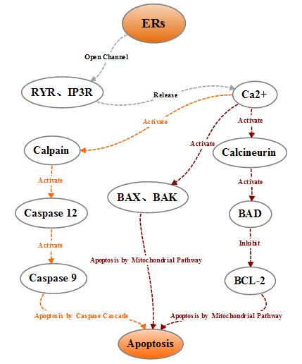
2.2.2 Regulation of apoptosis by the imbalance of Ca2+ homeostasis
During normal operation of the cells, endoplasmic reticulum releases Ca2 + in the endoplasmic reticulum into the cytoplasm mainly through the RyR and IP3R channels, and pumps intracellular Ca2 + into the endoplasmic reticulum lumen through a calcium pump to maintain the endoplasmic reticulum Ca2 + homeostasis. When the endoplasmic reticulum receives the stress signal, the Ca2 + homeostasis in the endoplasmic reticulum is broken, a large amount of Ca2 + enters the intracellular and mitochondria, which on the one hand influences the activity of mitochondria and Bcl-2 family proteins and leads the cells to apoptosis, on the other hand activates the intracellular neutral cysteine endopeptidase Calpain, the activated Calpain can activate caspase cascade and affect apoptosis.
Click for more apoptotic antibodies
Past review
Apoptosisi mediated by mitochondria
The next notice
Apoptosisi mediated by death receptor
3. Cited References
[1] Green D R, Kroemer G. The pathophysiology of mitochondrial cell death [J]. Science, 2004, 305: 626-629. Groenendyk J, Michalak M. Endoplasmic reticulum quality control and apoptosis [J]. Acta Biochimica Polonica, 2005, 52(2): 381-395.
[2] Bastida-Ruiz D, Aguilar E, Ditisheim A, et al. Endoplasmic reticulum stress responses in placentation - A true balancing act [J]. Placenta, 2017, 57: 163-169.
[3] Kang-sheng LIU, Zheng-hang PENG, Weng-jun CHENG, et al. Endoplasmic reticulum stress-induced apoptosis in the development of reproduction [J]. Reproductive and Developmental Medicine, 2016, 27(1): 51-59.
[4] Li J, Lee B, Lee A S. Endoplasmic reticulum stress-induced apoptosis: multiple pathways and activation of p53-up-regulated modulator of apoptosis (PUMA) and NOXA by p53 [J]. Journal of Biological Chemistry, 2006, 281(11): 7260-7270.
[5] Burton G J, Yung H W, Murray A J. Mitochondrial - Endoplasmic reticulum interactions in the trophoblast: Stress and senescence [J]. Placenta, 2017, 52: 146-155.
[6] Marchi S, Patergnani S, Missiroli S, et al. Mitochondrial and endoplasmic reticulum calcium homeostasis and cell death [J]. Cell Calcium, 2017.
[7] Breckenridge D G, Germain M, Mathai J P, et al. Regulation of apoptosis by endoplasmic reticulum pathways [J]. Oncogene, 2003, 22(53): 8608-8618.


Comments
Leave a Comment