Rat glial fibrillary acidic protein,GFAP ELISA Kit

Code CSB-E08602r
Size 96T,5×96T,10×96T
Price Request a Quote or Start an on-line Chat
Trial Size 24T ELISA Kit Trial Size (Only USD$150/ kit)
* Sample kit cost can be deducted as a $30 credit for each 96-assay kit of the same analyte and brand you subsequently purchase in six months till depleted. Apply now

Product Details

Target Name
glial fibrillary acidic protein
Alternative Names
Gfap ELISA Kit; Glial fibrillary acidic protein ELISA Kit; GFAP ELISA Kit
Abbreviation
Uniprot No.
Species
Rattus norvegicus (Rat)
Sample Types
serum, plasma, tissue homogenates
Detection Range
78 pg/mL-5000 pg/mL
Sensitivity
19.5 pg/mL
Assay Time
1-5h
Sample Volume
50-100ul
Detection Wavelength
450 nm
Research Area
Neuroscience
Assay Principle
quantitative
Measurement
Sandwich
Precision
Intra-assay Precision (Precision within an assay): CV%<8%      
Three samples of known concentration were tested twenty times on one plate to assess.  
Inter-assay Precision (Precision between assays): CV%<10%      
Three samples of known concentration were tested in twenty assays to assess.    
             
Linearity
To assess the linearity of the assay, samples were spiked with high concentrations of rat GFAP in various matrices and diluted with the Sample Diluent to produce samples with values within the dynamic range of the assay.
  Sample Serum(n=4)  
1:1 Average % 92  
Range % 88-98  
1:2 Average % 99  
Range % 94-109  
1:4 Average % 95  
Range % 89-98  
1:8 Average % 98  
Range % 89-104  
Recovery
The recovery of rat GFAP spiked to levels throughout the range of the assay in various matrices was evaluated. Samples were diluted prior to assay as directed in the Sample Preparation section.
Sample Type Average % Recovery Range  
Serum (n=5) 104 98-108  
EDTA plasma (n=4) 98 94-106  
             
             
Typical Data
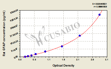
These standard curves are provided for demonstration only. A standard curve should be generated for each set of samples assayed.
pg/ml OD1 OD2 Average Corrected  
5000 2.804 2.925 2.865 2.664  
2500 2.123 2.201 2.162 1.961  
1250 1.529 1.507 1.518 1.317  
625 0.936 0.914 0.925 0.724  
312 0.590 0.576 0.583 0.382  
156 0.424 0.432 0.428 0.227  
78 0.309 0.300 0.305 0.104  
0 0.204 0.198 0.201    
Troubleshooting
and FAQs
Storage
Store at 2-8°C. Please refer to protocol.
Shelf Life
6 months
Lead Time
3-5 working days after you place the order, and it takes another 3-5 days for delivery via DHL or FedEx.
Description

This Rat GFAP ELISA Kit was designed for the quantitative measurement of Rat GFAP protein in serum, plasma, tissue homogenates. It is a Sandwich ELISA kit, its detection range is 78 pg/mL-5000 pg/mL and the sensitivity is 19.5 pg/mL .

Customer Reviews and Q&A

 Customer Reviews

There are currently no reviews for this product.

Submit a Review here

Target Background

Function
(From Uniprot)
GFAP, a class-III intermediate filament, is a cell-specific marker that, during the development of the central nervous system, distinguishes astrocytes from other glial cells.
Gene References into Functions
  1. demonstrate increased DNA methylation coupled with decreased histone acetylation at the Gfap promoter leading to suppression of Gfap expression under maternal hypothyroidism PMID: 29852171
  2. The present study found that social isolation during adolescence resulted in abnormal locomotor, emotional and cognitive behaviors and increased the expression of GFAP, ANXA2 and VIM in PFC of adult rats PMID: 28705472
  3. GFAP-positive structures were present and exhibited a tendency to become linear on both sides, with an increased density on the left. NFAP-positive expression was present in the left treated limb with a disorganized pattern PMID: 28652433
  4. We found that GFAP exhibited enhanced stability upon the addition of two equivalents of each ligands with ceftriaxone imparting a more spontaneous interactions and a more ordered complex system than phenytoin PMID: 27133445
  5. reduction of GFAP+ cell density was in agreement with a lower expression of GFAP protein PMID: 27579183
  6. Astrocyte expression of GLAST/GFAP was reduced via JAK1/JAK2/STAT3 signaling pathway after exposure to sevoflurane. PMID: 27003918
  7. Upon ependymal stem cells differentiation, Cx50 expression favors glial cell fate, since higher expression levels, endogenous or by over-expression of Cx50, augmented the expression of the astrocyte marker GFAP and impaired the neuronal marker Tuj1. PMID: 26561800
  8. It is likely that a plastic change in GFAP expression in astrocytes selectively occurs around Oxytocin (OXT) neurons at proestrus and facilitates OXT release. PMID: 26994384
  9. investigated temporal profile of astrocytic and neuronal injury markers after TBI; different mechanisms underlie clearance of UCH-L1 and GFAP in CSF and serum PMID: 25763798
  10. Its antibody is able to protect cells from oxidative stress, which is due to changed protein expressions of the actin cytoskeleton. PMID: 25837926
  11. GFAP release in hippocampus is significantly increased in a model of traumatic brain injury. PMID: 25898931
  12. Hippocampal glucose uptake defects correlate with NeuN immunoreactivity in the latent phase and GFAP immunoreactivity in the chronic phase. PMID: 25798055
  13. paracrine factors inhibit p38 MAPK and JNK, and most likely by regulating their downstream targets, p53 and STAT1, to promote astrocyte survival associated with GFAP downregulation after ischemic stroke in vitro PMID: 25752945
  14. The down-regulation of histone deacetylase SIRT1 abrogating the effect of cocoa on glial fibrillary acidic protein up-regulation and on Lys310-RelA/p65 acetylation by silencing or blockage was clearly demonstrated herein in vivo and in vitro. PMID: 25448608
  15. Data indicate that glial fibrillary acidic protein (GFAP) was up-regulated in satellite glial cells (SGCs) in dorsal root ganglia 14 days after streptozotocin injection. PMID: 25312986
  16. increased in nucleus ambiguous after recurrent or superior laryngeal nerve injury PMID: 25181319
  17. Diametric expression of GFAP and a different morphological pattern of caspase-3 labelling, although no changes in the cell number, were observed in the neurons of young and old animals. PMID: 25182537
  18. Progesterone promotes neuroprotection following traumatic brain injury by inhibiting the expression of Nogo-A and GFAP, and increasing GAP-43 expression. PMID: 24567055
  19. GFAP expression study also showed that cortical layer I usually contained multiple large astrocytes with branching processes, as well as numerous smaller processes with high intensity of expression. PMID: 25282817
  20. GFAP repression is mediated through direct binding of p-PPARgamma (S112) to its promoter region. PMID: 24481447
  21. GFAP is an important marker in determining the severity of traumatic brain injury. PMID: 24379073
  22. To clarify whether GFAP-positive neoplastic astrocytes exist in rat spontaneous oligodendrogliomas and mixed gliomas or not, immunohistochemical examination was performed on spontaneous oligodendrogliomas (26 cases) and mixed gliomas. PMID: 23076037
  23. Findings indicate the reciprocal relationships between GFAP expression and astrocyte neurotrophic activity by linking the ERalpha to ERbeta ratio to GFAP expression. PMID: 23515288
  24. TMJ inflammation induced significant upregulation of GFAP (an astroglial marker) in the trigeminal subnucleus caudalis. PMID: 23110394
  25. Western blot analysis showed that the chronic stress downregulated GFAP but upregulated NDRG2 protein PMID: 22610521
  26. In the hippocampus, thalamus, and piriform cortex both vascular laminin and astrocytic GFAP expression are upregulated following kainic acid injection. PMID: 22475395
  27. Increases in c-fos and GFAP were triggered by the combined stress of non-thermal irradiation and the toxic effect of picrotoxin on cerebral tissues. PMID: 21524663
  28. There was a reduction in GFAP levels in the hippocampus of non-trained diabetic animals, which was not found in trained diabetic group. PMID: 21892662
  29. Zuogui Pill may have protective effects on the optic nerve and retina ganglion cells after contusion by promoting nestin and GFAP expressions in Muller cells of the retina. PMID: 21906524
  30. Activation of a pro-survival IL-6/JAK2/STAT3 cascade contributes to cholera toxin-induced GFAP expression. PMID: 21470923
  31. Cesarean section might increase GFAP expression in the hippocampus and frontal cortex, and trigger neuronal apoptosis of hippocampus region. PMID: 21211284
  32. GFAP-positive temporomandibular joint disc cells were characterized by broader processes and existed exclusively in the deeper area. PMID: 21679183
  33. GFAP expression was down-regulated while GAP-43 expression upregulated in the retinal ganglial cells after peripheral nerve transplantation. PMID: 20423852
  34. Region-specific alterations in the gene expression of GFAP, VEGF, and FGF-2 and their receptors in the aged brain correspond to changes in astrocytic reactivity, supporting astrocytic heterogeneity and demonstrating a differential aging effect. PMID: 21385309
  35. GFAP content was diminished after a long-term administration of progesterone in hippocampus. PMID: 21683086
  36. There is a differential expression pattern of GFAP in the rat brain during pregnancy and the beginning of lactation that is associated with changes in brain function during these reproductive stages. PMID: 20732387
  37. Data show that astrocyte structural proteins GFAP and vimentin are induced by chronic leptin administration, suggesting astrocytes participating in leptin modulated synaptic inputs. PMID: 21343257
  38. Butylphthalide may protect the neuron-vascular unit of the hippocampus of Alzheimer model rats by inhibiting the expression of GFAP and increasing the expression of VEGF. PMID: 20197608
  39. Pax3 negatively regulates GFAP expression during astrocyte differentiation in vitro PMID: 21371476
  40. these results indicate that intracellular cAMP levels regulate the expression of NPP1 in rat C6 glioma cells by a signalling pathway that is different from the GFAP signal transduction pathway PMID: 21168404
  41. hyperbaric oxygenation treatment for cerebral infarction, can increase the expression of Map-2 and decrease the expression of GFAP. PMID: 19141977
  42. Amniotic fluid-GFAP levels appear to correlate with spinal cord injury as gestation proceeds in fetal rats with myelomeningocele. PMID: 21284970
  43. Retinal injury induces an upregulation of a complement of four intermediate filament proteins, including synemin and nestin, in Muller cells. PMID: 21139996
  44. Chondroitinase ABC can reduce the expression of GFAP after spinal cord injury. PMID: 21046809
  45. retinoic acid and cytokines have a synergistic effect on the regulation of glial fibrillary acidic protein expression PMID: 20876578
  46. There was no significant increase in GFAP immunoreactivity in the white matter in a weight drop model of mild cerebral contusion injury in the rat. PMID: 20479526
  47. Alcohol could repress the expression of GFAP and S100 of astrocytes. PMID: 16986516
  48. S100 protein and glial fibrillary acidic protein expression increased significantly in the hippocampal astrocytes of rats with Alzheimer disease, and were inhibited by butylphthalide. PMID: 19726345
  49. Brain concussion induced the expression of GFAP. PMID: 16850587
  50. expression of 25-kDa synaptosomal-associated protein and glial fibrillary acidic protein, were significantly enhanced in the cerebellum of rats with treadmill training after cerebral infarct PMID: 19661770

Show More

Hide All

Subcellular Location
Cytoplasm.
Protein Families
Intermediate filament family
Tissue Specificity
Expressed in the cortex and hippocampus. Expression decreases following acute and chronic corticosterone treatment.
Database Links
icon of phone
Call us
301-363-4651 (Available 9 a.m. to 5 p.m. CST from Monday to Friday)
icon of address
Address
7505 Fannin St., Ste 610, Room 7 (CUBIO Innovation Center), Houston, TX 77054, USA
icon of social media
Join us with

Subscribe newsletter

Leave a message

* To protect against spam, please pass the CAPTCHA test below.
CAPTCHA verification
© 2007-2024 CUSABIO TECHNOLOGY LLC All rights reserved. 鄂ICP备15011166号-1