Code | CSB-MP733578HU |
MSDS | |
Size | $228 |
Order now | |
Image |
|
Have Questions? | Leave a Message or Start an on-line Chat |
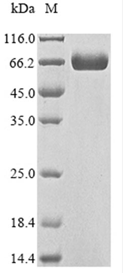
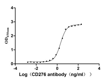
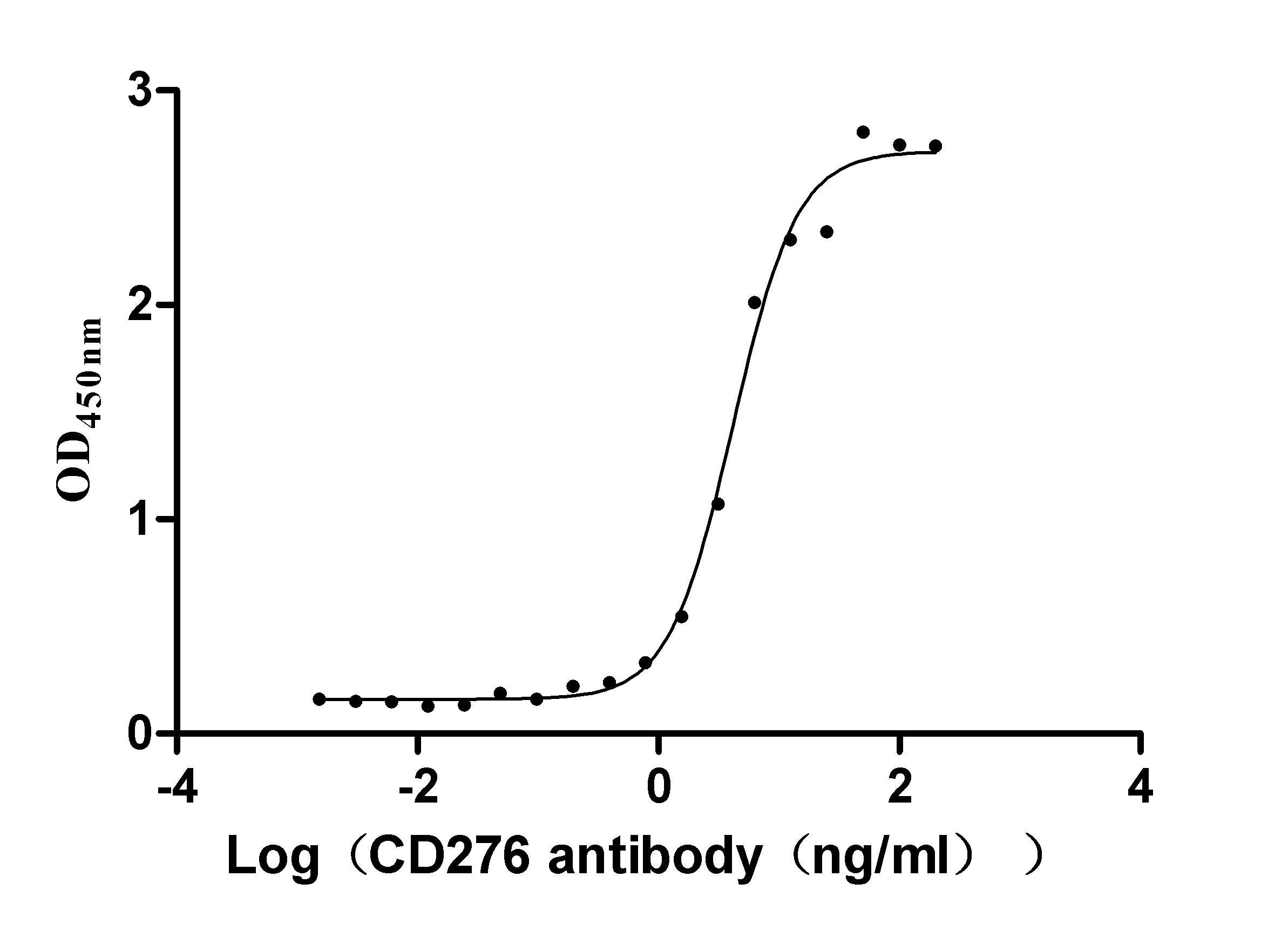
The DNA fragment corresponding to Leu29-Gly245 of the human CD276 antigen is fused with an FC-Myc-tag at the C-terminus and then is expressed in the mammalian cells. The resulting product is the recombinant human CD276 antigen. Its purity is over 90% measured by SDS-PAGE. On the gel, this protein was migrated to the molecular weight band of about 60-80 kDa. The endotoxin content of this CD276 protein is less than 1.0 EU/ug as determined by the LAL method. Its bioactivity has been validated through an ELISA. In the functional ELISA, this recombinant CD276 protein can bind to the anti-CD276 antibody, and the EC50 is 1.961-2.243 ng/ml. This CD276 protein is available now.
CD276 is an immune checkpoint molecule in the epithelial-mesenchymal transition (EMT) pathway. It is involved in cell proliferation, invasion, and metastasis in malignancies. Overexpression of CD276 has been found in numerous solid tumors such as breast, colorectal, and lung cancer, and has been strongly linked to a poor clinical prognosis.
Applications : Antigen, ELISA standard and its raw materials
Review: I used the CSB-MP733578HU protein for an ELISA experiment, and the EC50 was measured to be 0.3038-0.6364 ng/ml. This antigen has been validated by ELISA experiment and has good activity, making it suitable for later detection of animal immune serum titers and screening of hybridoma-positive clones.
By Anonymous
Are CUSABIO recombinant proteins sterile?
Yes, we can offer an aseptic processing service and it is free of charge, but you should remark this information when placing the order. We've performed aseptic processing for liquid protein before lyophilization, but there may exist contamination during lyophilization process. To clarify, lyophilized proteins can’t guarantee aseptic processing for the whole manufacture process.
Can you remove the endotoxin?
Not all endotoxin can be removed. Please contact us in advance if you need to remove the tag which takes 2-3 business days. We could offer endotoxin removal service free of charge using PMB affinity chromatography, use LAL reagent to semi-quantitatively detect the content of endotoxin, and guarantee endotoxin level within 0.1 ng/μg (1 EU/μg).
How to avoid inclusion bodies and improve soluble expression?
a. For proteins with high hydrophobicity or transmembrane domains, you should add fusion tags or add heat shock chaperones, and induce for a shorter time at low temperature or change to poor media. Generate truncated forms of protein or use membrane-rich strains.
b. For incorrect disulfide bond formation, you should add fusion partners, including thioredoxin, DsbA, DsbC. Clone in a vector containing secretion signal peptide to cell periplasm. Use gamiB (DE3)strains with the oxidative cytoplasmic environment. Lower inducer concentration and induction temperature.
c. For incorrect folding, you should use a fusion partner. Co-express with molecular chaperones. Use strains with cold-adapted chaperones. Supplement media with chemical chaperones and cofactors. Reduce the inducer concentration and add fresh media. Induce for a shorter time at low temperature.
Earlier studies found that B7H3 (also known as B7H3) promotes the activation of T cells. Chapoval et al. confirmed that in the presence of anti-CD3 antibodies, B7H3 can promote the proliferation of CD4 and CD8+ T cells and selectively promote the secretion of IFN-γ. And B7H3 transfection into tumor cells can enhance the killing ability of CTL. Further research found that only TLT-2 transgenic cells could bind to mouse B7H3 with high affinity, and TLT-2 was determined to be the receptor molecule of B7H3. Moreover, Hashiguchi et al. confirmed that the B7H3-TLT-2 pathway enhanced T cell activation. However, Leitner et al. did not find the specific binding of B7H3 to TLT-2 by flow cytometry, therefore, the exact receptor molecule of B7H3 is still unclear.
On the other hand, studies have found that B7H3 can also suppress T-cell immune responses. Some studies have shown that B7H3 can inhibit human and mouse T cells by activating or inhibiting NFTA (nuclear factor for activated T cells), NF-KB (nuclear factor kB) and AP-1 (activator protein-1) pathways. activation. In addition, results have demonstrated that B7H3 may inhibit T cell immune responses by inhibiting the activity of Thl. The study of Leiner et al. also found that B7H3 can down-regulate the secretion of IL-2 in T cells to inhibit the activity of T cells.