Call us
301-363-4651 (Available 9 a.m. to 5 p.m. CST from Monday to Friday)
| Code | CSB-MA878942A1m |
| Size | US$210 |
| Order now | |
| Image |
|
| Have Questions? | Leave a Message or Start an on-line Chat |
| Application | Recommended Dilution |
|---|---|
| WB | 1:1000-1:5000 |
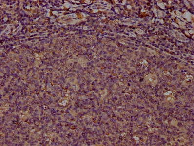
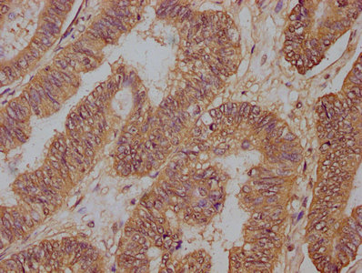
| IHC | 1:50-1:200 |
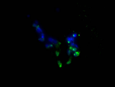
| IF | 1:50-1:200 |
In the preparation of this PD-L1 monoclonal antibody, a mouse is selected as the host animal. The recombinant human PD-L1 protein (19-238aa) was injected into the mouse to induce an immune response. Next, the spleen cells were removed from the immunized mouse and then fused with myeloma cells to generate hybridomas. PD-L1 antibody-producing-hybridomas were selected to culture. The PD-L1 monoclonal antibody was extracted from the mouse ascites and underwent purification by protein G affinity chromatography. Its specificity for human PD-L1 protein was validated in multiple applications, including ELISA, WB, IHC, IF, and FC.
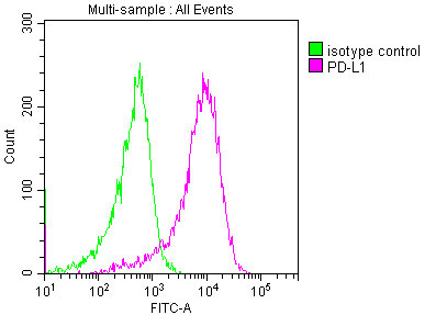
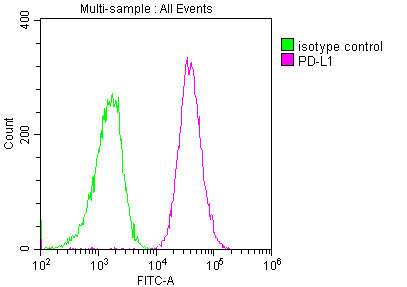
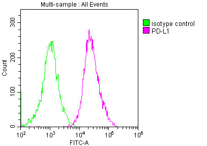
PD-L1 plays a key role in regulating the immune system. When PD-L1 binds to its receptor PD-1 on T cells, it inhibits the T cell response and prevents it from attacking PD-L1-expressing cells. This mechanism is often exploited by cancer cells, which can upregulate PD-L1 expression to evade destruction by the immune system. Targeting PD-L1 with immunotherapy drugs has become an important treatment strategy for some types of cancer.
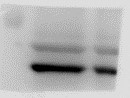
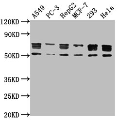
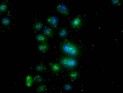
Applications : Western Blot (WB)
Sample type: Human granulosa cell carcinoma KGN
Sample dilution: 1:1500
Review: The antibody strip is correct, but there is a weak band nearby. The technician suggests that experiment steps are to be optimized.
By Anonymous
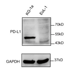
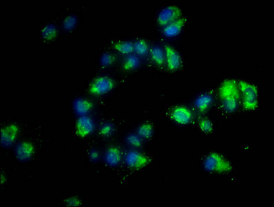
Applications : Western Blot (WB)
Review: To investigate whether PD-L1 expression is involved in the biological activities of leukemic cells, siRNA knockdown of PD-L1 expression in KG-1a via western blot analysis showed high expression of PD-L1.
By Anonymous

