CD274 Antibody

Code CSB-PA878942ESR1HU
Size US$166
Order now
Image
  • Immunohistochemistry of paraffin-embedded human colon tissue using CSB-PA878942ESR1HUat dilution of 1:100

Have Questions? Leave a Message or Start an on-line Chat

Product Details

Full Product Name
Rabbit anti-Homo sapiens (Human) CD274 Polyclonal antibody
Uniprot No.
Target Names
CD274
Alternative Names
B7 H antibody; B7 H1 antibody; B7 homolog 1 antibody; B7-H1 antibody; B7H antibody; B7H1 antibody; CD 274 antibody; CD274 antibody; CD274 antigen antibody; CD274 molecule antibody; MGC142294 antibody; MGC142296 antibody; OTTHUMP00000021029 antibody; PD L1 antibody; PD-L1 antibody; PD1L1_HUMAN antibody; PDCD1 ligand 1 antibody; PDCD1L1 antibody; PDCD1LG1 antibody; PDL 1 antibody; PDL1 antibody; Programmed cell death 1 ligand 1 antibody; Programmed death ligand 1 antibody; RGD1566211 antibody
Raised in
Rabbit
Species Reactivity
Human
Immunogen
Recombinant Human Programmed cell death 1 ligand 1 protein (19-238AA)
Immunogen Species
Homo sapiens (Human)
Conjugate
Non-conjugated
Clonality
Polyclonal
Isotype
IgG
Purification Method
Antigen Affinity Purified
Concentration
It differs from different batches. Please contact us to confirm it.
Buffer
PBS with 0.02% sodium azide, 50% glycerol, pH7.3.
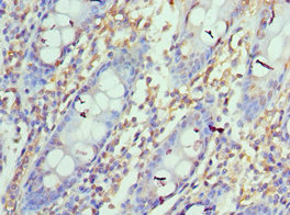
Form
Liquid
Tested Applications
ELISA, IHC
Recommended Dilution
Application Recommended Dilution
IHC 1:20-1:200
Troubleshooting and FAQs
Storage
Upon receipt, store at -20°C or -80°C. Avoid repeated freeze.
Lead Time
Basically, we can dispatch the products out in 1-3 working days after receiving your orders. Delivery time maybe differs from different purchasing way or location, please kindly consult your local distributors for specific delivery time.

Customer Reviews and Q&A

 Customer Reviews

There are currently no reviews for this product.

Submit a Review here

Target Background

Function
Plays a critical role in induction and maintenance of immune tolerance to self. As a ligand for the inhibitory receptor PDCD1/PD-1, modulates the activation threshold of T-cells and limits T-cell effector response. Through a yet unknown activating receptor, may costimulate T-cell subsets that predominantly produce interleukin-10 (IL10).; The PDCD1-mediated inhibitory pathway is exploited by tumors to attenuate anti-tumor immunity and escape destruction by the immune system, thereby facilitating tumor survival. The interaction with PDCD1/PD-1 inhibits cytotoxic T lymphocytes (CTLs) effector function. The blockage of the PDCD1-mediated pathway results in the reversal of the exhausted T-cell phenotype and the normalization of the anti-tumor response, providing a rationale for cancer immunotherapy.
Gene References into Functions
  1. an HBV-pSTAT3-SALL4-miR-200c axis regulates PD-L1 causing T cell exhaustion PMID: 29593314
  2. hypothesized that an oncolytic poxvirus would attract T cells into the tumour, and induce PD-L1 expression in cancer and immune cells, leading to more susceptible targets for anti-PD-L1 immunotherapy PMID: 28345650
  3. multivariate Cox hazards regression analysis identified ALCAM and PD-L1 (both P < 0.01) as potential independent risk factors for primary diffuse pleural mesotheliomas PMID: 28811252
  4. miR-191-5p was identified to have negative correlation with PD-L1 expression and acted as an independent prognostic factor of OS in patients with colon adenocarcinoma. PMID: 30045644
  5. our findings suggest a regulatory mechanism of PD-L1 through data analysis, in vitro and vivo experiments, which is an important factor of immune evasion in GC cells, and CXCL9/10/11-CXCR3 could regulate PD-L1 expression through STAT and PI3K-Akt signaling pathways in GC cells. PMID: 29690901
  6. CD163(+)CD204(+) Tumor-associated macrophages possibly play a key role in the invasion and metastasis of oral squamous cell carcinoma by T-cell regulation via IL-10 and PD-L1 production. PMID: 28496107
  7. Results demonstrated that the expression of PDL1 in colorectal carcinoma tissue was significantly increased compared with the paracancerous tissue. Blocking PDL1 can inhibit tumor growth by activating CD4+ and CD8+ T cells involved in the immune response. PMID: 30272332
  8. miR-574-3p was identified to potentially regulate PD-L1 expression in chordoma, which inversely correlated with PD-L1. Positive PD-L1 expression on tumor cells was associated with advanced stages and TILs infiltration, whereas decreased miR-574-3p level correlated with higher muscle invasion, more severe tumor necrosis and poor patient survival. PMID: 29051990
  9. PD-L1 expression was detected in 69% of cases of primary melanoma of the vulva PMID: 28914674
  10. PD-L1 tumor cell expression is strongly associated with increased HIF-2alpha expression and presence of dense lymphocytic infiltration in clear cell renal cell carcinoma. PMID: 30144808
  11. Different Signaling Pathways in Regulating PD-L1 Expression in EGFR Mutated Lung Adenocarcinoma PMID: 30454551
  12. PD-L1, Ki-67, and p53 staining individually had significant prognostic value for patients with stage II and III colorectal cancer PMID: 28782638
  13. Challenging PD-L1 expressing cytotoxic T cells as a predictor for response to immunotherapy in melanoma PMID: 30050132
  14. Our results confirm and extend prior studies of PD-L1 and provide new data of PD-L2 expression in lymphomas PMID: 29122656
  15. Positive PD-L1 expression is indicative of worse clinical outcome in Xp11.2 renal cell carcinoma. PMID: 28522811
  16. PD-L1 expression in cancer cells is upregulated in response to DNA double-strand break. PMID: 29170499
  17. Targeting PD-L1 Protein is an efficient anti-cancer immunotherapy strategy. (Review) PMID: 30264678
  18. Suggest that PD-L1 may play a relevant role in metastatic spread and may be a candidate prognostic biomarker in cutaneous squamous cell carcinoma. PMID: 29742559
  19. PD-L1 immunostaining scoring for non-small cell lung cancer based on immunosurveillance parameters. PMID: 29874226
  20. SLC18A1 might complement other biomarkers currently under study in relation to programmed cell death protein 1/programmed cell death protein ligand 1 inhibition PMID: 30194079
  21. Low PDL1 expression is associated with mammary and extra-mammary Paget disease. PMID: 29943071
  22. Low PDL1 mRNA expression is associated with non-muscle-invasive bladder cancer. PMID: 29150702
  23. we found that in advanced stage NSCLC patients who received nivolumab, the C allele of PD-L1 rs4143815 and the G allele of rs2282055 were significantly associated with better ORR and PFS. This is the first report that PD-L1 SNP, which was thought to increase PD-L1 expression, is associated with a response to nivolumab. PMID: 28332580
  24. PD-L1 expression differs between the two components of lung ASCs. Given the complexity of lung ASCs, their treatment outcomes may be improved by administration of both EGFR TKIs and anti-PD-1/PD-L1 antibodies in cases where EGFR mutations are present and PD-L1 is overexpressed. PMID: 28387300
  25. IDO and B7-H1 expressions were observed in patients with pancreatic carcinoma tissues and are important markers for PC malignant progression PMID: 30029936
  26. There was higher programmed cell death protein ligand-1(PD-L1) expression in post-treatment EBV DNA-positive patients. Post-treatment positive EBV DNA status maybe a useful biomarker of worse outcomes in early stage -stage extranodal natural killer/T cell lymphoma. PMID: 30116872
  27. PD-L1 is a critical TTP-regulated factor that contributes to inhibiting antitumor immunity. PMID: 29936792
  28. Structural and functional analyses unexpectedly reveal an N-terminal loop outside the IgV domain of PD-1. This loop is not involved in recognition of PD-L1 but dominates binding to nivolumab, whereas N-glycosylation is not involved in binding at all. PMID: 28165004
  29. While mutational analysis appeared similar to that of older patients with OCSCC who lack a smoking history, a comparatively high degree of PD-L1 expression and PD-1/L1 concordance (P=0.001) was found among young female OCSCC patients. PMID: 28969885
  30. PD-L1 expression is predictive of survival in diffuse large B-cell lymphoma, irrespective of rituximab treatment. PMID: 29748856
  31. PD-L1 expression was augmented on CD8+ T cells in BALF of a patient with smoldering adult T-cell lymphoma and Pneumocystis jiroveci pneumonia. This suggested that the PD-1-PD-L1 system may suppress not only antitumor immunity but also host defense against pathogens and thereby allow establishment of chronic HTLV-1 infection and immunodeficiency. PMID: 28967040
  32. MUC1 drives PD-L1 expression in triple-negative breast cancer cells. PMID: 29263152
  33. Positive PD-L1 expression was found in 36.8% of inflammatory breast carcinoma (IBC) samples but was not significantly associated with the clinicopathologic variables examined. Worse overall survival (OS) was significantly associated with positive PD-L1, negative estrogen receptor, and triple-negative status. The 5-year OS rate was 36.4% for patients with PD-L1-positive IBC and 47.3% for those with PD-L1-negative. PMID: 29425258
  34. PD-L1 expression display a highly variable distribution in clear cell renal cell carcinomas and this particularity should be kept in mind when selecting the tumor samples to be tested for immunotherapy. PMID: 29661736
  35. Report relatively low level PD-L1 positivity in treatment-naive acinar prostatic adenocarcinoma. PMID: 30257853
  36. High PD-L1 expression is associated with pulmonary metastases in head and neck squamous cell carcinoma. PMID: 29937180
  37. these data suggest that DNA-damage signaling is insufficient for upregulating PD-L1 in normal human dermal fibroblasts PMID: 29859207
  38. High CD274 expression is associated with Oral Squamous Cell Carcinoma. PMID: 28669079
  39. This study provides important evidence of higher levels of agreement of PD-L1 expression in pulmonary metastasis compared with in multiple primary lung cancer, and high positivity of PD-L1 expression in pulmonary metastatic lesions with wild-type EGFR in an Asian population. PMID: 29254651
  40. High PD-L1 expression is associated with Mycobacterium avium complex-induced lung disease. PMID: 28169347
  41. PD-L1 expression in tumor-associated immune cells may be associated with a higher probability of clinical response to avelumab in metastatic breast cancer PMID: 29063313
  42. The expression of PDL1 was significantly increased following treatment with gefitinib. PMID: 29901173
  43. These results demonstrated that the IFNGinduced immunosuppressive properties of B7H1 in human BM and WJMSCs were mediated by STAT1 signaling, and not by PI3K/RACalpha serine/threonineprotein kinase signaling PMID: 29901104
  44. PD-1/PD-L1 expression is a frequent occurrence in poorly differentiated neuroendocrine carcinomas of the digestive system. PMID: 29037958
  45. High CD274 expression is associated with Epithelial Ovarian Cancer. PMID: 30275195
  46. Studied expression levels of CD274 molecule (PD-L1) in thymic epithelial tumors. Found PD-L1 expression level correlated with the degree of TET malignancy. PMID: 29850538
  47. Authors assessed PD-L1 expression in both tumor cells and tumor-infiltrating immune cells in the tumor specimens (complete histological sections, not tissue microarray). PMID: 28420659
  48. We conclude that a subgroup of advanced disease ovarian cancer patients with high grade tumors, expressing PD-L1, may be prime candidates for immunotherapy targeting PD-1 signaling PMID: 29843813
  49. PD-L1 expression is a prognostic factor related with poor survival among patients that developed non-small cell lung cancer. PMID: 29614306
  50. The inhibition of PTEN also reduced the cancer effects of CD4+ T cells on non-small cell lung cancer (NSCLC) cell lines following miR-142-5p downregulation. Therefore, our study demonstrated that miR-142-5p regulated CD4+ T cells in human NSCLC through PD-L1 expression via the PTEN pathway. PMID: 29767245

Show More

Hide All

Subcellular Location
Cell membrane; Single-pass type I membrane protein. Early endosome membrane; Single-pass type I membrane protein. Recycling endosome membrane; Single-pass type I membrane protein.; [Isoform 1]: Cell membrane; Single-pass type I membrane protein.; [Isoform 2]: Endomembrane system; Single-pass type I membrane protein.
Protein Families
Immunoglobulin superfamily, BTN/MOG family
Tissue Specificity
Highly expressed in the heart, skeletal muscle, placenta and lung. Weakly expressed in the thymus, spleen, kidney and liver. Expressed on activated T- and B-cells, dendritic cells, keratinocytes and monocytes.
Database Links

HGNC: 17635

OMIM: 605402

KEGG: hsa:29126

STRING: 9606.ENSP00000370989

UniGene: Hs.521989

CUSABIO guaranteed quality
icon of phone
Call us
301-363-4651 (Available 9 a.m. to 5 p.m. CST from Monday to Friday)
icon of address
Address
7505 Fannin St., Ste 610, Room 7 (CUBIO Innovation Center), Houston, TX 77054, USA
icon of social media
Join us with

Subscribe newsletter

Leave a message

* To protect against spam, please pass the CAPTCHA test below.
CAPTCHA verification
© 2007-2024 CUSABIO TECHNOLOGY LLC All rights reserved. 鄂ICP备15011166号-1
Place an order now

I. Product details

*
*
*
*

II. Contact details

*
*

III. Ship To

*
*
*
*
*
*
*

IV. Bill To

*
*
*
*
*
*
*
*