Code | CSB-EP004950HU1 |
Size |
US$2466Purchase it in Cusabio online store (only available for customers from the US) |
Image | |
Have Questions? | Leave a Message or Start an on-line Chat |
Description |
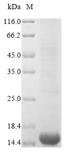
The DNA coding sequence translated into the human CD63 protein sequence (103-203aa) was fused with the N-terminal 6xHis tag sequence to form the recombinant DNA, which was inserted into an expression vector. The reconstructed expression vector was transformed into the E.coli for follow-up expression. The product underwent purification to obtain the recombinant human CD63 protein with N-terminal 6xHis tag. The SDS-PAGE analysis determined its purity higher than 85%. After electrophoresis, a 15 kDa protein band was observed on the gel. CD63 is a gene encoding a protein named CD63 antigen (also known as LAMP-3 or Tspan-30) in human and belongs to Tetraspanin (TM4SF) family. CD63 is mainly associated with membranes of intracellular vesicles. A study has reported exosomal markers CD63 and CD9 are elevated in pancreatic tumor tissues. In this study, authors have indicated that CD63 is a critical player in LMP1 exosomal trafficking and LMP1-mediated enhancement of exosome production and may play further roles in limiting downstream LMP1 signaling. Furthermore, emerging evidence demonstrated CD63 tetraspanin slows down cell migration and translocates to the endosomal-lysosomal-MIICs route after extracellular stimuli in human immature dendritic cells. |
Purity | Greater than 85% as determined by SDS-PAGE. |
Target Names | CD63 |
Uniprot No. | P08962 |
Research Area | Cancer |
Alternative Names | CD63; MLA1; TSPAN30; CD63 antigen; Granulophysin; Lysosomal-associated membrane protein 3; LAMP-3; Lysosome integral membrane protein 1; Limp1; Melanoma-associated antigen ME491; OMA81H; Ocular melanoma-associated antigen; Tetraspanin-30; Tspan-30; CD antigen CD63 |
Species | Homo sapiens (Human) |
Source | E.coli |
Expression Region | 103-203aa |
Target Protein Sequence | AGYVFRDKVMSEFNNNFRQQMENYPKNNHTASILDRMQADFKCCGAANYTDWEKIPSMSKNRVPDSCCINVTVGCGINFNEKAIHKEGCVEKIGGWLRKNV Note: The complete sequence including tag sequence, target protein sequence and linker sequence could be provided upon request. |
Mol. Weight | 15.5 kDa |
Protein Length | Partial |
Tag Info |
N-terminal 6xHis-tagged |
Form |
Liquid or Lyophilized powder Note: We will preferentially ship the format that we have in stock, however, if you have any special requirement for the format, please remark your requirement when placing the order, we will prepare according to your demand. |
Buffer | If the delivery form is liquid, the default storage buffer is Tris/PBS-based buffer, 5%-50% glycerol.
Note: If you have any special requirement for the glycerol content, please remark when you place the order. If the delivery form is lyophilized powder, the buffer before lyophilization is Tris/PBS-based buffer, 6% Trehalose, pH 8.0. |
Reconstitution | We recommend that this vial be briefly centrifuged prior to opening to bring the contents to the bottom. Please reconstitute protein in deionized sterile water to a concentration of 0.1-1.0 mg/mL.We recommend to add 5-50% of glycerol (final concentration) and aliquot for long-term storage at -20°C/-80°C. Our default final concentration of glycerol is 50%. Customers could use it as reference. |
Troubleshooting and FAQs |
Protein FAQs |
Storage Condition | Store at -20°C/-80°C upon receipt, aliquoting is necessary for mutiple use. Avoid repeated freeze-thaw cycles. |
Shelf Life | The shelf life is related to many factors, storage state, buffer ingredients, storage temperature
and the stability of the protein itself. Generally, the shelf life of liquid form is 6 months at -20°C/-80°C. The shelf life of lyophilized form is 12 months at -20°C/-80°C. |
Lead Time | 3-7 business days |
Notes | Repeated freezing and thawing is not recommended. Store working aliquots at 4°C for up to one week. |
Datasheet & COA | Please contact us to get it. |
Applications : control proteins
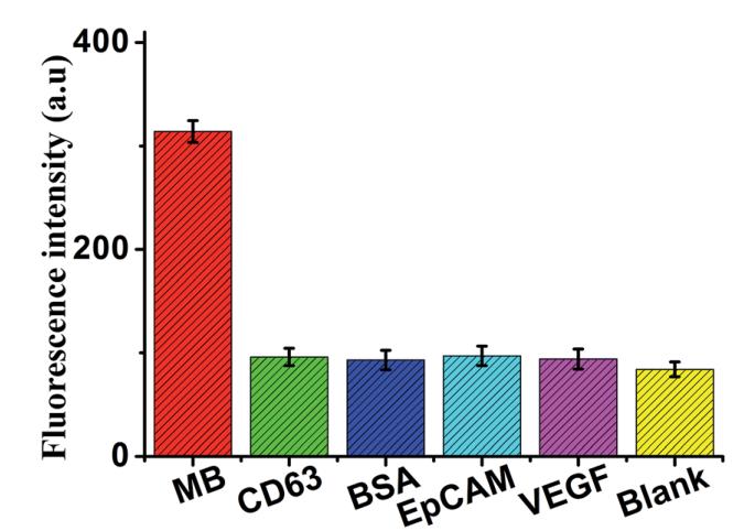
Review: Fluorescence intensity (at the emission wavelength of 517.6 nm) of the sensor in the presence of MB (5 ng mL -1 ), CD63 (50 ng mL -1 ), BSA (50 ng mL -1 ), EPCAM (50 ng mL -1 ), VEGF (50 ng mL -1 ) and black, respectively. Error bars: SD, n = 3.
By Anonymous
Function |
Functions as cell surface receptor for TIMP1 and plays a role in the activation of cellular signaling cascades. Plays a role in the activation of ITGB1 and integrin signaling, leading to the activation of AKT, FAK/PTK2 and MAP kinases. Promotes cell survival, reorganization of the actin cytoskeleton, cell adhesion, spreading and migration, via its role in the activation of AKT and FAK/PTK2. Plays a role in VEGFA signaling via its role in regulating the internalization of KDR/VEGFR2. Plays a role in intracellular vesicular transport processes, and is required for normal trafficking of the PMEL luminal domain that is essential for the development and maturation of melanocytes. Plays a role in the adhesion of leukocytes onto endothelial cells via its role in the regulation of SELP trafficking. May play a role in mast cell degranulation in response to Ms4a2/FceRI stimulation, but not in mast cell degranulation in response to other stimuli.
|
Gene References into Functions |
|
Subcellular Location | Cell membrane; Multi-pass membrane protein. Lysosome membrane; Multi-pass membrane protein. Late endosome membrane; Multi-pass membrane protein. Endosome, multivesicular body. Melanosome. Secreted, extracellular exosome. Cell surface. |
Protein Families | Tetraspanin (TM4SF) family |
Tissue Specificity | Detected in platelets (at protein level). Dysplastic nevi, radial growth phase primary melanomas, hematopoietic cells, tissue macrophages. |
Database Links |
HGNC: 1692 OMIM: 155740 KEGG: hsa:967 STRING: 9606.ENSP00000257857 UniGene: Hs.445570 |