Call us
301-363-4651 (Available 9 a.m. to 5 p.m. CST from Monday to Friday)

Host Cell Proteins (HCP) are residual protein impurities derived from host cells (such as yeast, E. coli, mammalian cells, insect cells, etc.) during the production of biopharmaceuticals. These impurities may accompany the target drug protein during cell culture, fermentation, or expression. Despite purification processes, they may still remain in the final product, potentially affecting the safety and efficacy of the drug.
The potential risks posed by HCP residues include:
Accurate measurement of HCP residue levels is not only critical for ensuring clinical drug safety but also serves as a cornerstone for optimizing production processes, reducing R&D risks, and ensuring compliance with global regulatory standards.
| Method | Technical Features | Applications |
|---|---|---|
| ELISA (Enzyme-Linked Immunosorbent Assay) |
|
|
| Mass Spectrometry (LC-MS/MS) |
|
|
| Western Blot |
|
|
| Two-Dimensional Gel Electrophoresis (2D-PAGE) |
|
|
| Capillary Electrophoresis (CE) |
|
|
| HPLC |
|
|
As the industry "gold standard," ELISA is widely recognized by global regulatory agencies for its simplicity, high sensitivity, and reliable results. Leveraging a mature enzyme-linked immunosorbent assay platform, CUSABIO has developed a series of HCP detection kits suitable for various host cells. These kits enable precise quantification of HCP residues in production stages (cell culture supernatants, purification intermediates, final products) for expression systems such as CHO, HEK293, and E. coli, providing reliable analytical tools for biopharmaceutical process development and quality control.
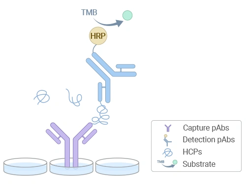
Schematic diagram of HCP residue detection by ELISA
High Sensitivity
Uses high-titer antibodies (titer ≥106) combined with affinity purification, achieving detection limits as low as ng/mL.
Broad Antibody Coverage
Employs high-affinity polyclonal antibodies to recognize most host cell proteins, ensuring comprehensive detection.
High Stability
Incorporates broad-spectrum protein stabilizers and microplate processing technology, minimizing intra- and inter-batch variability.
Strong Compatibility
Optimized buffer system reduces interference from complex matrices.
End-to-End Solution
Covers detection across all production stages, compliant with global regulatory standards.
For different types of test samples, different detection kits are set up. Please click on the product details page to select the appropriate kit based on your experimental needs.
| Code | Product Name |
|---|---|
| CSB-EQ33270CHO | Chinese Hamster Ovary (CHO) Host Cell Protein (HCP) ELISA Kit(Supernatant) |
| CSB-EQ33269HEK | 293-related cell line (293T, 293F)Host Cell Protein (HCP) ELISA Kit |
| CSB-EQ33268SF | Sf9 host cell protein (HCP) ELISA Kit |
| CSB-EQ33267OP | Ogataea Polymorpha host cell protein (HCP) ELISA Kit |
| CSB-EQ33266PY | Pichia Yeast GS115 host cell protein (HCP) ELISA Kit |
| CSB-EQ33260ECO | E. coli Host Cell Protein (HCP) ELISA Kit(3 Strains) |
| CSB-EQ33259ECO | E. coli Host Cell Protein (HCP) ELISA Kit(6 Strains) |
| CSB-EQ33265PY | Pichia Yeast X-33 host cell protein (HCP) ELISA Kit |
| CSB-EQ33264HEK | 293 Host Cell Protein (HCP) ELISA Kit(Intracellular&Supernatant) |
| CSB-EQ33263CHO | Chinese Hamster Ovary (CHO) Host Cell Protein (HCP) ELISA Kit(Supernatant) |
| CSB-EQ33262CHO | Chinese Hamster Ovary (CHO) Host Cell Protein (HCP) ELISA Kit(Intracellular) |
| CSB-EQ33261ECO | E. coli Host Cell Protein (HCP) ELISA Kit(3 Strains) |
Q1: What sample types are compatible with this kit?
A: This kit can quantitatively detect HCP residues in culture supernatants, protein purification intermediates, and final products. It is suitable for biopharmaceutical R&D, process optimization, and quality control.
Q2: What detection principle does this kit use?
A: The kit employs a double-antibody sandwich method, forming an "antibody-antigen-antibody" complex with coated antibodies and HRP-labeled detection antibodies. HCP residue levels are quantified via TMB color development and OD measurement.
Q3: What are the storage conditions for kit components? Can opened reagents be reused?
A: All reagents (excluding the sealing film) must be stored sealed at 2–8 °C. Unused microplate strips may be resealed with the sealing film and returned to the foil bag for storage at 4 °C. All reagents and components should be used within one week after opening.
Q4: Why is spike recovery testing necessary? How is it performed?
A: The spike recovery experiment is used to evaluate whether the sample matrix interferes with the detection results. For example, for product CSB-EQ33263CHO, it is recommended to mix diluted standard S1 (400 ng/mL) with the test sample at a ratio of 1:3 (such as adding 25 μL of standard S1 with or without a concentration of 400 ng/mL into 75 μL of test solution). The recovery rate should then be calculated. An ideal recovery rate should fall within 80–120% to ensure data reliability.

