Mouse glial fibrillary acidic protein,GFAP ELISA Kit

Code CSB-E08603m
Size 96T,5×96T,10×96T
Price Request a Quote or Start an on-line Chat
Trial Size 24T ELISA Kit Trial Size (Only USD$150/ kit)
* Sample kit cost can be deducted as a $30 credit for each 96-assay kit of the same analyte and brand you subsequently purchase in six months till depleted. Apply now

Product Details

Target Name
glial fibrillary acidic protein
Alternative Names
GfapGlial fibrillary acidic protein ELISA Kit; GFAP ELISA Kit
Abbreviation
Uniprot No.
Species
Mus musculus (Mouse)
Sample Types
serum, plasma, tissue homogenates
Detection Range
3.12 pg/mL-200 pg/mL
Sensitivity
0.78 pg/mL
Assay Time
1-5h
Sample Volume
50-100ul
Detection Wavelength
450 nm
Research Area
Neuroscience
Assay Principle
quantitative
Measurement
Sandwich
Precision
Intra-assay Precision (Precision within an assay): CV%<8%
Three samples of known concentration were tested twenty times on one plate to assess.
Inter-assay Precision (Precision between assays): CV%<10%
Three samples of known concentration were tested in twenty assays to assess.
Linearity
To assess the linearity of the assay, samples were spiked with high concentrations of mouse GFAP in various matrices and diluted with the Sample Diluent to produce samples with values within the dynamic range of the assay.
SampleSerum(n=4)
1:1Average %90
Range %85-94
1:2Average %98
Range %95-101
1:4Average %102
Range %97-108
1:8Average %94
Range %91-97
Recovery
The recovery of mouse GFAP spiked to levels throughout the range of the assay in various matrices was evaluated. Samples were diluted prior to assay as directed in the Sample Preparation section.
Sample TypeAverage % RecoveryRange
Serum (n=5) 9791-104
EDTA plasma (n=4)9390-96
Typical Data
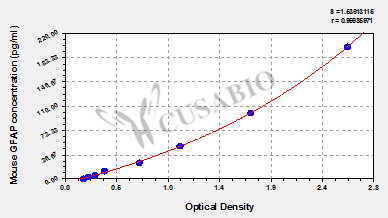
These standard curves are provided for demonstration only. A standard curve should be generated for each set of samples assayed.
pg/mlOD1OD2AverageCorrected
2002.563 2.595 2.579 2.400
1001.636 1.764 1.700 1.521
501.027 1.095 1.061 0.882
250.704 0.667 0.686 0.507
12.50.387 0.359 0.373 0.194
6.250.286 0.284 0.285 0.106
3.120.233 0.222 0.228 0.049
00.173 0.184 0.179
Troubleshooting
and FAQs
Storage
Store at 2-8°C. Please refer to protocol.
Shelf Life
6 months
Lead Time
3-5 working days after you place the order, and it takes another 3-5 days for delivery via DHL or FedEx.
Description

This Mouse GFAP ELISA Kit was designed for the quantitative measurement of Mouse GFAP protein in serum, plasma, tissue homogenates. It is a Sandwich ELISA kit, its detection range is 3.12 pg/mL-200 pg/mL and the sensitivity is 0.78 pg/mL.

Citations

Customer Reviews and Q&A

 Customer Reviews
Average Rating:
5.0 - 1 reviews

Submit a Review here

Sample type: Plasma (anticoagulant)

Sample species: Mouse

Review: I used to detect GFAP from Mouse plasma

By Anonymous

Target Background

Function
(From Uniprot)
GFAP, a class-III intermediate filament, is a cell-specific marker that, during the development of the central nervous system, distinguishes astrocytes from other glial cells.
Gene References into Functions
  1. This report the successful prediction and validation of Gfap as an miR-3099 target gene using a combination of bioinformatics resources with enrichment of annotations based on functional ontologies and a spatio-temporal expression dataset. PMID: 28597341
  2. These results indicate that autoantibodies against GFAP could serve as a predictive marker for the development of overt autoimmune diabetes. PMID: 28546444
  3. GFAP is specifically expressed in the auricular chondrocytes, and assumes a pivotal role in resistance against mechanical stress. PMID: 28063220
  4. compared open-skull and thinned-skull imaging methods for two-photon laser microscopy of live astrocytes in neocortex of GFAP-GFP transgenic mice PMID: 28107381
  5. work reveals that an Alexander disease-causing mutation alters GFAP turnover kinetics in vivo and provides an essential foundation for future studies aimed at preventing or reducing the accumulation of GFAP. PMID: 28223355
  6. Tat expression or GFAP expression led to formation of GFAP aggregates and induction of unfolded protein response (UPR) and endoplasmic reticulum (ER) stress in astrocytes. PMID: 27609520
  7. Study provides evidence that transcription of one of the astrocyte-specific genes, Gfap, is cooperatively regulated by co-expressed genes and their regulatory factors. PMID: 27041678
  8. This study demonstrated the GFAP-ApoE4 mice exhibited motor impairments when compared to GFAP-ApoE3 and wild-type mice. PMID: 26892275
  9. PINK1 deficiency causes defects in GFAP-positive astrogliogenesis during brain development. PMID: 26746235
  10. Gnasxl deficiency does not directly affect glial development in the hypothalamus, since it is expressed in neurons, and Gfap-positive astrocytes and tanycytes appear normal during early postnatal stages. PMID: 27080240
  11. Induction of glial cytokine expression was sequential, aligned with active sickness behavior, and preceded increased Iba-1 or GFAP immunoreactivity after lipopolysaccharide challenge PMID: 26470014
  12. Study provides a mechanistic link between the GFAP mutations/overexpression and the symptoms in those affected with Type II Alexander disease PMID: 26190408
  13. Study described GFAP-expressing non-myelinating Schwann cells in the lung, validated a transgenic mouse line that drives expression of cre under a GFAP promoter PMID: 26442852
  14. findings thus show that the inability to produce GFAP and Vim affects normal retinal physiology and that the effect of IF deficiency on retinal cell survival differs, depending on the underlying pathologic condition PMID: 26251181
  15. CUL4B as a negative regulator of GFAP expression during neural development. PMID: 26025376
  16. Astrocytes deficient of GFAP and vimentin showed decreased Notch signal sending competence and altered expression of Notch signaling pathway-related genes PMID: 26118771
  17. Absence of GFAP, or both GFAP and vimentin, alters Alzheimer's disease-induced changes in gene expression profile of astrocytes, showing a compensation of the decrease of neuronal support genes and a trend for a higher inflammatory expression profile PMID: 25731615
  18. Data indicate that glial fibrillary acidic protein (GFAP) was up-regulated in satellite glial cells (SGCs) in dorsal root ganglia 14 days after streptozotocin injection. PMID: 25312986
  19. Findings demonstrate that ENT1 regulates GFAP expression and possibly astrocyte function PMID: 25365803
  20. Data suggest that prenatal alterations in expression of various fetal brain proteins (including down-regulation of Gfap) are associated with aberrant behavioral characteristics of transgenic mice that model autism-like behavior. PMID: 25849768
  21. Study shows that increased levels of astrocytic GFAP can contribute to increases on inter-ictal spikes but do not represent a risk factor for appearance of post-traumatic seizures, even when there is increase in reactive gliosis PMID: 25069089
  22. The Phactr4 signals were not associated with F-actin fibers but were closely associated with intermediate filaments such as nestin and glial fibrillary acidic protein (GFAP) fibers. PMID: 24748504
  23. Report increasing glial fibrillary acidic protein expression and electron microscopic features of brain edema in rodent cerebral malaria. PMID: 24966914
  24. absence of GFAP and vimentin in glial cells does not seem to affect the outcome after peripheral motoneuron injury but may have an important effect on the response dynamics PMID: 24223940
  25. the astrocyte became activated, exhibiting significantly increased levels of GFAP expression directly related to the level of HIV/VSV replication PMID: 24254728
  26. traumatic scratch injury to astrocytes triggered a calcium influx from the extracellular compartment and activated the JNK/c-Jun/AP-1 pathway to switch on GFAP PMID: 24123203
  27. Data show that CD8 T cells reactive to glial fibrillary acidic protein (GFAP), a protein expressed in astrocytes, drive unique aspects of inflammatory central nervous system autoimmunity. PMID: 24591371
  28. Brain levels of GFAP and Tau proteins decreased significantly at 6 h and increased considerably at 24 h after repeated blast exposures. Plasma samples showed a similar initial decrease and later increase over this timeframe. PMID: 23933206
  29. Data indicate that Gfapdelta is expressed in the in developing mouse brain sub-ventricular zones in accordance with the described localization in the developing and adult human brain. PMID: 23991052
  30. GFAP was found to be downregulated in HSV-1 acute infection in cornea and upregulated in late stage, suggesting that GFAP might play some role during HSV-1 infection in cornea. PMID: 23758602
  31. Data show that neurofibromatosis type 1 (NF1)-inactivation results in a cell-autonomous increase in glial fibrillary acidic protein+ (GFAP+), but not in NG2 proteoglycan NG2+, cell proliferation in vitro. PMID: 23318450
  32. this study demonistrated that mouse models of Alexander disease exhibit significant pathology in GFAP-positive radial glia-like cells in the dentate gyrus, and suffer from deficits in adult neurogenesis. PMID: 24259590
  33. These studies demonstrate that transactivation of the Gfap promoter is an early and sustained indicator of the disease process in the mouse. PMID: 23432455
  34. These data suggest that all astroglia cells in the developing and adolescent mouse brain express GFAPdelta, regardless of their neurogenic capabilities. PMID: 23285135
  35. The GFAP-stained intensity of the retinal area is increased in contralateral eyes and decreased in retinal ganglion cells of eyes with laser-induced ocular hypertension. PMID: 22583833
  36. Postulate that glial cells with increased Gfap expression support the elongation of new neurites from retinal ganglion cells possibly by providing a scaffold for outgrowth. PMID: 23259929
  37. GFAP expression is almost not affected by melatonin treatment in aged mice. PMID: 22200709
  38. differential regulation of GFAP isoforms is not involved in the reorganization of the intermediate filament network in reactive gliosis or in neurogenesis in the mouse brain. PMID: 22912745
  39. CD44-positive cells are APCs in the early postnatal cerebellum; surviving cells gradually express glial fibrillary acidic protein GFAP), a marker for mature astrocytes, indicating differentiation into mature astrocytes is the default for these cells. PMID: 21732075
  40. GFAP-negative astrocytes are fully inflammation-competent, displaying phenotypic heterogeneity as is commonly observed in brain astrocytes. PMID: 22072312
  41. In a mouse model of amyotrophic lateral sclerosis (ALS), GFAP is not necessary for the initiation of disease; instead, it plays some modulatory roles in the progression of ALS. PMID: 21453731
  42. Treadmill exercise training decreased the expression of GFAP in the striatum of chronic Parkinsonian mice, which can partially explain the beneficial neuroprotective role of exercise in patients with parkinson disease. PMID: 21725169
  43. The study shows opposing pattern of nestin and glial fibrillary acidic protein expression in mouse hippocampus occuring in early postnatal development, suggesting that it is important for neural differentiation and positioning in the hippocampus. PMID: 21368556
  44. In the mouse there is a slight increase in the number of GFAP positive cells in the white matter after 3 days of severe cerebral contusion trauma. PMID: 20479526
  45. Increased expression of GFAP in Muller cells of mer knockout mice occur at P20d in the peripheral retina and P4w in the central retina. GFAP expression in Muller cells appears to be a secondary response to the loss of retinal neurons. PMID: 20497693
  46. One protein, GFAP (glial fibrillary acidic protein), was found to be elevated in the LINCL mice compared with normal controls in both isolated storage bodies and a lysosome-enriched subcellular fraction that contains storage material. PMID: 20370715
  47. The results of this study suggested that GFAP is necessary for morphological retention and distribution of reactive astrocytes during prion disease, and that there is a GFAP-dependent function of glial filaments in reactive astrocytes. PMID: 19931516
  48. Report demonstrated for the first time that GFAPdelta is specifically expressed in radial glia and SVZ neural progenitors during human brain development. PMID: 20040497
  49. The exact expression of glial fibrillary acidic protein (GFAP) in trigeminal ganglion and dental pulp. PMID: 11838710
  50. Human influenza viral infection in utero alters GFAP immunoreactivity in the developing brains of neonatal mice. PMID: 12140787

Show More

Hide All

Subcellular Location
Cytoplasm.
Protein Families
Intermediate filament family
Tissue Specificity
Brain; isoform 2 expressed at 20-fold lower level than isoform 1.
Database Links
icon of phone
Call us
301-363-4651 (Available 9 a.m. to 5 p.m. CST from Monday to Friday)
icon of address
Address
7505 Fannin St., Ste 610, Room 7 (CUBIO Innovation Center), Houston, TX 77054, USA
icon of social media
Join us with

Subscribe newsletter

Leave a message

* To protect against spam, please pass the CAPTCHA test below.
CAPTCHA verification
© 2007-2024 CUSABIO TECHNOLOGY LLC All rights reserved. 鄂ICP备15011166号-1