Call us
301-363-4651 (Available 9 a.m. to 5 p.m. CST from Monday to Friday)
| Code | CSB-EP330493CYZ |
| Abbreviation | Recombinant Campylobacter fetus sapA protein, partial |
| MSDS | |
| Size | $388 |
| Order now | |
| Image |
|
| Have Questions? | Leave a Message or Start an on-line Chat |
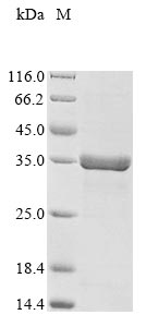
Synthesizing the recombinant Campylobacter fetus S-layer protein (sapA) generally involves integrating the DNA fragment that encodes the Campylobacter fetus sapA protein (1-268aa) into a plasmid along with the N-terminal 10xHis-tag and C-terminal Myc-tag gene, introducing the recombinant plasmid into E.coli cells, followed by the selection and culturing of positive E.coli cells, induction of protein expression, and subsequent cell lysis. The protein is purified through affinity purification, and SDS-PAGE analysis is conducted to confirm the presence of the protein and determine its purity. The protein's purity surpasses 85%.
Saposin A is a glycosphingolipid activator protein that is crucial in breaking down cerebroside sulfates within the lysosome [1]. It is derived from the proteolytic processing of domain 1 of its precursor protein, prosaposin [2]. Saposin A, along with other saposins like B, C, and D, acts at the lipid-water interface in lysosomes, facilitating the hydrolysis of membrane components by water-soluble exohydrolases [3]. Saposin A has tissue-specific effects on glycosphingolipid degradation, as observed in mutant mice studies [4]. Additionally, saposin A has been linked to neurological deficits and glycosphingolipid accumulation in cases of saposin B deficiency [5].
Saposin A is part of a family of saposins, each with distinct functions. Saposin B, for example, is involved in the activation of arylsulfatase A, β-galactosidase, α-galactosidase, and neuraminidase [6]. On the other hand, saposin C acts as an activator of the lysosomal enzyme glucocerebrosidase, which hydrolyzes glucocerebroside into glucose and cerebroside [7]. The crystal structures of saposins A and C have been elucidated, showing compact monomeric structures with small hydrophobic cores [8][9].
References:
[1] V. Ahn, K. Faull, J. Whitelegge, A. Fluharty, & G. Privé, Crystal structure of saposin b reveals a dimeric shell for lipid binding, Proceedings of the National Academy of Sciences, vol. 100, no. 1, p. 38-43, 2002. https://doi.org/10.1073/pnas.0136947100
[2] S. Morimoto, B. Martin, Y. Yamamoto, K. Kretz, J. O’Brien, & Y. Kishimoto, Saposin a: second cerebrosidase activator protein., Proceedings of the National Academy of Sciences, vol. 86, no. 9, p. 3389-3393, 1989. https://doi.org/10.1073/pnas.86.9.3389
[3] R. Schultz-Heienbrok, N. Remmel, R. Klingenstein, M. Rossocha, K. Sandhoff, & T. Maier, Crystallization and preliminary characterization of three different crystal forms of human saposin c heterologously expressed inpichia pastoris, Acta Crystallographica Section F Structural Biology and Crystallization Communications, vol. 62, no. 2, p. 117-120, 2006. https://doi.org/10.1107/s1744309105043186
[4] Y. Sun, M. Zamzow, H. Ran, W. Zhang, B. Quinn, S. Barneset al., Tissue-specific effects of saposin a and saposin b on glycosphingolipid degradation in mutant mice, Human Molecular Genetics, vol. 22, no. 12, p. 2435-2450, 2013. https://doi.org/10.1093/hmg/ddt096
[5] Y. Sun, D. Witte, H. Ran, M. Zamzow, S. Barnes, H. Chenget al., Neurological deficits and glycosphingolipid accumulation in saposin b deficient mice, Human Molecular Genetics, vol. 17, no. 15, p. 2345-2356, 2008. https://doi.org/10.1093/hmg/ddn135
[6] M. Champagne, S. Lamontagne, & M. Potier, Binding of gm1‐ganglioside to a synthetic peptide derived from the lysosomal sphingolipid‐activator‐protein saposin b, Febs Letters, vol. 347, no. 2-3, p. 265-267, 1994. https://doi.org/10.1016/0014-5793(94)00536-2
[7] E. Alba, S. Weiler, & N. Tjandra, Solution structure of human saposin c: ph-dependent interaction with phospholipid vesicles, Biochemistry, vol. 42, no. 50, p. 14729-14740, 2003. https://doi.org/10.1021/bi0301338
[8] V. Ahn, P. Leyko, J. Alattia, L. Chen, & G. Privé, Crystal structures of saposins a and c, Protein Science, vol. 15, no. 8, p. 1849-1857, 2006. https://doi.org/10.1110/ps.062256606
[9] K. Popovic and G. Privé, Structures of the human ceramide activator protein saposin d, Acta Crystallographica Section D Biological Crystallography, vol. 64, no. 5, p. 589-594, 2008. https://doi.org/10.1107/s0907444908003120
There are currently no reviews for this product.

