Recombinant Canine parvovirus type 2 Capsid protein VP2, partial

In Stock
Code CSB-EP351680CCR
Abbreviation Recombinant Canine parvovirus type 2 Capsid protein VP2 protein, partial
MSDS
Size $388
Order now
Image
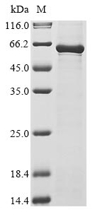
  • (Tris-Glycine gel) Discontinuous SDS-PAGE (reduced) with 5% enrichment gel and 15% separation gel.
Have Questions? Leave a Message or Start an on-line Chat

Product Details

Purity
Greater than 85% as determined by SDS-PAGE.
Target Names
N/A
Uniprot No.
Research Area
others
Alternative Names
Coat protein VP2
Species
Canine parvovirus type 2 (CPV-2)
Source
E.coli
Expression Region
30-553aa
Target Protein Sequence
GGGGGSGGVGISTGTFNNQTEFKFLENGWVEITANSSRLVHLNMPESENYRRVVVNNMDKTAVNGNMALDDIHVQIVTPWSLVDANAWGVWFNPGDWQLIVNTMSELHLVSFEQEIFNVVLKTVSESATQPPTKVYNNDLTASLMVALDSNNTMPFTPAAMRSETLGFYPWKPTIPTPWRYYFQWDRTLVPSHTGTSGTPTNIYHGTDPDDVQFYTIENSVPVHLLRTGDEFATGTFFFDCKPCRLTHTWQTNRALGLPPFLNSLPQSEGATNFGDIGVQQDKRRGVTQMGNTNYITEATIMRPAEVGYSAPYYSFEASTQGPFKTPIAAGRGGAQTYENQAADGDPRYAFGRQHGQKTTTTGETPDRITYIAHHDTGRYPEGDWIQNINFNLPVTNDNVLLPTDPIGGKTGINYTNIFNTYGPLTALNNVPPVYPNGQIWDKEFDTDLKPRPHVNAPFVCQHNCPGQLFVKVAPNLTNEYDPDASANMSRIVTYSHFWWKGKLVFKAKLRASHTWNPIQQMSI
Note: The complete sequence may include tag sequence, target protein sequence, linker sequence and extra sequence that is translated with the protein sequence for the purpose(s) of secretion, stability, solubility, etc.
If the exact amino acid sequence of this recombinant protein is critical to your application, please explicitly request the full and complete sequence of this protein before ordering.
Mol. Weight
65.9 kDa
Protein Length
Partial
Tag Info
N-terminal 10xHis-tagged and C-terminal Myc-tagged
Form
Liquid or Lyophilized powder
Note: We will preferentially ship the format that we have in stock, however, if you have any special requirement for the format, please remark your requirement when placing the order, we will prepare according to your demand.
Buffer
If the delivery form is liquid, the default storage buffer is Tris/PBS-based buffer, 5%-50% glycerol. If the delivery form is lyophilized powder, the buffer before lyophilization is Tris/PBS-based buffer, 6% Trehalose.
Reconstitution
We recommend that this vial be briefly centrifuged prior to opening to bring the contents to the bottom. Please reconstitute protein in deionized sterile water to a concentration of 0.1-1.0 mg/mL.We recommend to add 5-50% of glycerol (final concentration) and aliquot for long-term storage at -20°C/-80°C. Our default final concentration of glycerol is 50%. Customers could use it as reference.
Troubleshooting and FAQs
Storage Condition
Store at -20°C/-80°C upon receipt, aliquoting is necessary for mutiple use. Avoid repeated freeze-thaw cycles.
Shelf Life
The shelf life is related to many factors, storage state, buffer ingredients, storage temperature and the stability of the protein itself.
Generally, the shelf life of liquid form is 6 months at -20°C/-80°C. The shelf life of lyophilized form is 12 months at -20°C/-80°C.
Lead Time
3-7 business days
Notes
Repeated freezing and thawing is not recommended. Store working aliquots at 4°C for up to one week.
Datasheet & COA
Please contact us to get it.
Description

The production of recombinant canine parvovirus type 2 Capsid protein VP2 begins with the isolation of the target gene corresponding to the 30-553aa of the canine parvovirus type 2 Capsid protein VP2. This gene is cloned into an expression vector with an N-terminal 10xHis-tag gene and a C-terminal Myc-tag gene. The expression vector is introduced into E. coli cells via transformation. The E. coli cells express the gene, producing the recombinant protein. This protein is harvested from the culture typically through cell lysis and purified using affinity chromatography. The final step involves measuring the purity of this recombinant protein, reaching up to 85%.

Canine parvovirus type 2 (CPV-2) is a significant pathogen that emerged in 1978, causing acute hemorrhagic enteritis and myocardial disease in young dogs [1]. The capsid protein VP2 is the main structural protein of CPV-2, responsible for viral entry and host cell recognition [2]. Research has shown that changes in specific amino acids in the VP2 capsid region have led to the divergence of CPV-2 into antigenic variants like CPV-2a and CPV-2b, affecting the virus's ability to infect hosts and adapt to different environments [3]. The VP2 protein of CPV-2 is crucial for inducing virus-neutralizing antibodies and determining the host range of the virus [4].

References:
[1] L. Shackelton, C. Parrish, U. Truyen, & E. Holmes, High rate of viral evolution associated with the emergence of carnivore parvovirus, Proceedings of the National Academy of Sciences, vol. 102, no. 2, p. 379-384, 2004. https://doi.org/10.1073/pnas.0406765102
[2] M. Vihinen‐Ranta, D. Wang, W. Weichert, & C. Parrish, The vp1 n-terminal sequence of canine parvovirus affects nuclear transport of capsids and efficient cell infection, Journal of Virology, vol. 76, no. 4, p. 1884-1891, 2002. https://doi.org/10.1128/jvi.76.4.1884-1891.2002
[3] N. Ahmed, A. Riaz, Z. Zubair, M. Saqib, S. Ijaz, M. Nawaz-ul-Rehmanet al., Molecular analysis of partial vp-2 gene amplified from rectal swab samples of diarrheic dogs in pakistan confirms the circulation of canine parvovirus genetic variant cpv-2a and detects sequences of feline panleukopenia virus (fpv), Virology Journal, vol. 15, no. 1, 2018. https://doi.org/10.1186/s12985-018-0958-y
[4] P. Zimmermann, M. Ritzmann, H. Selbitz, K. Heinritzi, & U. Truyen, Vp1 sequences of german porcine parvovirus isolates define two genetic lineages, Journal of General Virology, vol. 87, no. 2, p. 295-301, 2006. https://doi.org/10.1099/vir.0.81086-0

Customer Reviews and Q&A

 Customer Reviews

There are currently no reviews for this product.

Submit a Review here

Target Background

Function
Capsid protein self-assembles to form an icosahedral capsid with a T=1 symmetry, about 22 nm in diameter, and consisting of 60 copies of two size variants of the capsid proteins, VP1 and VP2, which differ by the presence of an N-terminal extension in the minor protein VP1. The capsid encapsulates the genomic ssDNA. Capsid proteins are responsible for the attachment to host cell receptor TFRC. This attachment induces virion internalization predominantly through clathrin-endocytosis. Binding to the host receptors also induces capsid rearrangements leading to surface exposure of VP1 N-terminus, specifically its phospholipase A2-like region and nuclear localization signal(s). VP1 N-terminus might serve as a lipolytic enzyme to breach the endosomal membrane during entry into host cell. Intracytoplasmic transport involves microtubules and interaction between capsid proteins and host dynein. Exposure of nuclear localization signal probably allows nuclear import of capsids.
Subcellular Location
Virion. Host nucleus.
Protein Families
Parvoviridae capsid protein family
icon of phone
Call us
301-363-4651 (Available 9 a.m. to 5 p.m. CST from Monday to Friday)
icon of address
Address
7505 Fannin St., Ste 610, Room 7 (CUBIO Innovation Center), Houston, TX 77054, USA
icon of social media
Join us with

Subscribe newsletter

Leave a message

* To protect against spam, please pass the CAPTCHA test below.
CAPTCHA verification
© 2007-2025 CUSABIO TECHNOLOGY LLC All rights reserved. 鄂ICP备15011166号-1
Place an order now

I. Product details

*
*
*
*

II. Contact details

*
*

III. Ship To

*
*
*
*
*
*
*

IV. Bill To

*
*
*
*
*
*
*
*