Recombinant Human Programmed cell death protein 6 (PDCD6)

In Stock
Code CSB-EP017672HU
Abbreviation Recombinant Human ALG2 protein
MSDS
Size US$212
Order now
Image
  • (Tris-Glycine gel) Discontinuous SDS-PAGE (reduced) with 5% enrichment gel and 15% separation gel.
Have Questions? Leave a Message or Start an on-line Chat

Product Details

Purity
Greater than 90% as determined by SDS-PAGE.
Target Names
PDCD6
Uniprot No.
Research Area
Apoptosis
Alternative Names
AIP1; ALG 2; ALG-2-interacting protein 1; ALG2; ALIX; Apoptosis linked gene 2; Apoptosis linked gene 2 protein; Apoptosis-linked gene 2 protein; FLJ42309; FLJ46208; Hp95; KIAA1375; MA 3; MA3; MGC111017; MGC119050; MGC9123; PDCD 6; Pdcd6; PDCD6_HUMAN; PEF 1B; PEF1B; Probable calcium binding protein ALG 2; Probable calcium binding protein ALG2; Probable calcium-binding protein ALG-2; Programmed cell death 6; Programmed cell death 6-interacting protein; Programmed cell death protein 6; PS 2; PS2
Species
Homo sapiens (Human)
Source
E.coli
Expression Region
1-191aa
Target Protein Sequence
MAAYSYRPGPGAGPGPAAGAALPDQSFLWNVFQRVDKDRSGVISDTELQQALSNGTWTPFNPVTVRSIISMFDRENKAGVNFSEFTGVWKYITDWQNVFRTYDRDNSGMIDKNELKQALSGFGYRLSDQFHDILIRKFDRQGRGQIAFDDFIQGCIVLQRLTDIFRRYDTDQDGWIQVSYEQYLSMVFSIV
Note: The complete sequence may include tag sequence, target protein sequence, linker sequence and extra sequence that is translated with the protein sequence for the purpose(s) of secretion, stability, solubility, etc.
If the exact amino acid sequence of this recombinant protein is critical to your application, please explicitly request the full and complete sequence of this protein before ordering.
Mol. Weight
48.9kDa
Protein Length
Full Length
Tag Info
N-terminal GST-tagged
Form
Liquid or Lyophilized powder
Note: We will preferentially ship the format that we have in stock, however, if you have any special requirement for the format, please remark your requirement when placing the order, we will prepare according to your demand.
Buffer
If the delivery form is liquid, the default storage buffer is Tris/PBS-based buffer, 5%-50% glycerol.
Note: If you have any special requirement for the glycerol content, please remark when you place the order.
If the delivery form is lyophilized powder, the buffer before lyophilization is Tris/PBS-based buffer, 6% Trehalose.
Reconstitution
We recommend that this vial be briefly centrifuged prior to opening to bring the contents to the bottom. Please reconstitute protein in deionized sterile water to a concentration of 0.1-1.0 mg/mL.We recommend to add 5-50% of glycerol (final concentration) and aliquot for long-term storage at -20°C/-80°C. Our default final concentration of glycerol is 50%. Customers could use it as reference.
Troubleshooting and FAQs
Storage Condition
Store at -20°C/-80°C upon receipt, aliquoting is necessary for mutiple use. Avoid repeated freeze-thaw cycles.
Shelf Life
The shelf life is related to many factors, storage state, buffer ingredients, storage temperature and the stability of the protein itself.
Generally, the shelf life of liquid form is 6 months at -20°C/-80°C. The shelf life of lyophilized form is 12 months at -20°C/-80°C.
Lead Time
3-7 business days
Notes
Repeated freezing and thawing is not recommended. Store working aliquots at 4°C for up to one week.
Datasheet & COA
Please contact us to get it.
Description

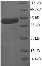
Recombinant Human Programmed cell death protein 6 (PDCD6) is produced in E. coli and contains the complete protein sequence from amino acids 1 to 191. It carries an N-terminal GST tag, which makes purification and detection more straightforward. The protein reaches purity levels above 90% as confirmed by SDS-PAGE, suggesting it meets high-quality standards for research work. This product is intended strictly for research use only and is not suitable for therapeutic or diagnostic applications.

PDCD6, also called ALG2, is a calcium-binding protein that appears to be involved in several cellular processes, including apoptosis and signal transduction pathways. It seems to play an important role in regulating programmed cell death, which is likely essential for maintaining cellular balance. Research on PDCD6 may be particularly significant because of its involvement in cellular stress responses and potential connections to apoptosis-related disorders.

Potential Applications

Note: The applications listed below are based on what we know about this protein's biological functions, published research, and experience from experts in the field. However, we haven't fully tested all of these applications ourselves yet. We'd recommend running some preliminary tests first to make sure they work for your specific research goals.

PDCD6 is a calcium-binding protein involved in apoptosis that requires proper folding and calcium-binding capability for its biological function. While E. coli can successfully express some eukaryotic proteins, the correct folding and functional activity cannot be guaranteed without validation. The large GST tag (≈26 kDa) may cause significant steric interference with the native protein structure (≈22 kDa), potentially disrupting functional domains. No validation data (e.g., calcium binding assays, apoptosis functional tests) are provided. Therefore, the protein's folding status and bioactivity remain unverified without verification.

1. Protein-Protein Interaction Studies

If correctly folded, the GST-tagged PDCD6 could be used for pull-down assays to identify binding partners, as the GST tag facilitates immobilization. However, if misfolded, interaction domains may be altered, leading to non-specific binding or failure to recognize genuine biological partners. The large GST tag may also sterically hinder proper protein-protein interactions, even with correct folding.

2. Antibody Development and Validation

This application is suitable, as antibody generation primarily relies on linear epitope recognition. The full-length protein provides comprehensive epitope coverage. However, antibodies generated against potentially misfolded proteins may not optimally recognize conformation-dependent epitopes of native PDCD6 in biological contexts.

3. Biochemical Characterization and Enzymatic Assays

If properly folded, the recombinant protein could be used for biochemical characterization. However, if misfolded, data on thermal stability and cofactor requirements would misrepresent the native protein's properties. The GST tag may dominate biophysical measurements, complicating the interpretation of PDCD6-specific characteristics.

4. ELISA-Based Quantitative Assays

The GST tag enables technical feasibility for ELISA development through anti-GST capture. However, if misfolded, the protein may not present native PDCD6 epitopes accurately, compromising assay specificity for detecting endogenous PDCD6. Results should be validated with native protein standards.

5. Structural and Biophysical Studies

If correctly folded, structural studies could provide valuable insights. However, the large GST tag would dominate structural signals and likely interfere with crystallization or NMR analysis of PDCD6-specific domains. If misfolded, any structural data would be biologically irrelevant.

Final Recommendation & Action Plan

This GST-tagged PDCD6 requires validation before functional applications. Recommended actions: (1) Remove GST tag using protease cleavage and purify tag-free PDCD6 for critical applications; (2) Validate folding through biophysical methods (circular dichroism, size-exclusion chromatography) and functional assays (calcium binding, apoptosis assays); (3) For interaction studies, include tag-free controls and validate findings with alternative methods; (4) For structural studies, consider expressing tag-free PDCD6 or using cleavable tag systems. Antibody development can proceed, but requires validation against native PDCD6. Always include proper controls and consider eukaryotic expression systems for functionally validated proteins.

Customer Reviews and Q&A

 Customer Reviews

There are currently no reviews for this product.

Submit a Review here

Target Background

Function
Calcium sensor that plays a key role in processes such as endoplasmic reticulum (ER)-Golgi vesicular transport, endosomal biogenesis or membrane repair. Acts as an adapter that bridges unrelated proteins or stabilizes weak protein-protein complexes in response to calcium: calcium-binding triggers exposure of apolar surface, promoting interaction with different sets of proteins thanks to 3 different hydrophobic pockets, leading to translocation to membranes. Involved in ER-Golgi transport by promoting the association between PDCD6IP and TSG101, thereby bridging together the ESCRT-III and ESCRT-I complexes. Together with PEF1, acts as calcium-dependent adapter for the BCR(KLHL12) complex, a complex involved in ER-Golgi transport by regulating the size of COPII coats. In response to cytosolic calcium increase, the heterodimer formed with PEF1 interacts with, and bridges together the BCR(KLHL12) complex and SEC31 (SEC31A or SEC31B), promoting monoubiquitination of SEC31 and subsequent collagen export, which is required for neural crest specification. Involved in the regulation of the distribution and function of MCOLN1 in the endosomal pathway. Promotes localization and polymerization of TFG at endoplasmic reticulum exit site. Required for T-cell receptor-, Fas-, and glucocorticoid-induced apoptosis. May mediate Ca(2+)-regulated signals along the death pathway: interaction with DAPK1 can accelerate apoptotic cell death by increasing caspase-3 activity. Its role in apoptosis may however be indirect, as suggested by knockout experiments. May inhibit KDR/VEGFR2-dependent angiogenesis; the function involves inhibition of VEGF-induced phosphorylation of the Akt signaling pathway. In case of infection by HIV-1 virus, indirectly inhibits HIV-1 production by affecting viral Gag expression and distribution.; Has a lower Ca(2+) affinity than isoform 1.
Gene References into Functions
  1. Up-regulation of miR-20a by HPV16 E6 exerted growth-promoting effects by targeting PDCD6 in cervical carcinoma cells. PMID: 29710555
  2. the t(5;21)(p15;q22) translocation could be identified only when what had seemed like a del(21)(qq) in G-banded preparations was examined using FISH and RNA-sequencing directed at finding out what lay behind the 21q-. PMID: 29672642
  3. ALG-2 binding to Scotin is strictly calcium dependent, indicating a role of this interaction in calcium signaling pathways PMID: 17889823
  4. found that targeting Requiem and Alg-2 did not result in extended culture viability, but resulted in an increase in maximum viable cell numbers and cumulative IVCD under fed-batch conditions PMID: 20571937
  5. the alg2 binding site is one of the key determinants of the retention kinetics of Sec31A at endoplasmic reticulum exit sites PMID: 20834162
  6. This is the first report showing interaction of ALG-2 with a P-body component (PATL1).PATL1 as well as DCP1A, a well-known P-body marker, co-localized with a subset of ALG-2. PMID: 22437941
  7. ALG-2 attenuates COPII budding in vitro and stabilizes the Sec23/Sec31A complex. PMID: 24069399
  8. Results show the crystal structure of the complex between ALG-2 and a peptide of Sec31A and found that the peptide binds to the third hydrophobic pocket (Pocket 3) and that ALG-2 recognizing 2 types of motifs at different hydrophobic surfaces of Sec31A. PMID: 25667979
  9. These results indicate that ALG-2 has an anti-apoptotic function in HeLa cells by facilitating the passage through checkpoints in the G2/M cell cycle phase. PMID: 19013425
  10. analysed the expression of ALG-2 in 7371 tumor tissue samples of various origin; most notably, ALG-2 was upregulated in mesenchymal tumors. PMID: 19383317
  11. ALG-2/Sec31A interactions were not required for the localization of Sec31A to ER exit sites per se but appeared to acutely regulate the stability and trafficking of the cargo receptor p24 and the distribution of the vesicle tether protein p115 PMID: 25006245
  12. Our study demonstrates that ALG-2 plays a role in the regulation of cytoskeletal rearrangement that drives cell polarization and migration in breast cancer cells. PMID: 27926525
  13. MAP1B heavy chain has a unique binding site for a calcium-binding protein ALG-2. PMID: 29432744
  14. These results suggest that a change in the intracellular calcium level plays a role in regulation of the secretory pathway via interaction of ALG-2 with MISSL and MAP1B. PMID: 28864773
  15. The gene copy numbers and mRNA levels for both ALG-2 and HEBP2 are significantly upregulated in breast and lung cancer. Coexpression of ALG-2 and HEBP2 markedly increases the cytoplasmic pool of ALG-2 and alters the subcellular distribution of HEBP2. Abnormalities in the ALG-2/HEBP2 interaction impairs spindle orientation and positioning during mitosis. PMID: 28004381
  16. The results suggest that ALG-2 acts as a Ca2+-sensitive adaptor to concentrate and polymerize TFG at endoplasmic reticulum exit sites, supporting a potential role for ALG-2 in COPII-dependent trafficking from the endoplasmic reticulum. PMID: 27813252
  17. ALG2 has a tumor suppressive role in glioblastoma and might be a potential target for the treatment of glioblastoma. PMID: 28300556
  18. miR-183 may function as an oncogene and may enhance cell proliferation by targeting PDCD6, implying a potential therapeutic target for this malignancy. PMID: 27738758
  19. Achieved results show that polymorphism rs3756712 is signifi cantly associated with the risk of endometriosis development in Slovak women. Polymorphism rs4957014 did not show any connection with development of endometriosis PMID: 27546697
  20. Our results suggest that PDCD6 may play an important role in the pathogenesis of cervical squamous cell carcinoma PMID: 25362542
  21. PDCD6 may represent a biomarker candidate gene that could help to identify a group of patients at high risk for recurrence and death. PMID: 24792888
  22. Palmitoylation sites and the N-terminal Pro-rich region were necessary for efficient secretion, but ABSs (ALG-2-binding sites) were dispensable. PMID: 23350699
  23. The present study provided evidence that rs4957014 and rs3756712 are associated with Uterine leiomyoma (UL) risk, the results indicated that genetic polymorphisms in PDCD6 may contribute to the development of UL. PMID: 23551056
  24. CHERP and ALG-2 participate in regulation of alternative splicing of IP3R1 pre-mRNA and provide new insights into post-transcriptional regulation of splicing variants in Ca(2+) signaling pathways. PMID: 24078636
  25. the binding of ALG-2 to IST1 is Ca(2+)-dependent PMID: 23649269
  26. the current study indicates that PDCD6 gene may be a new susceptibility gene to endometriosis. PMID: 23137875
  27. The results of this study show that rs4957014G/T is associated with an increased risk of non-small cell lung cancer (NSCLC), which suggest that PDCD6 gene single nucleotide polymorphism is a risk factor for susceptibility of NSCLC. PMID: 23167403
  28. we conclude that a novel p53-responsive gene PDCD6 is accumulated in the nucleus and induces apoptosis in response to DNA damage. PMID: 22712728
  29. PDCD6 seems to play an important role in ovarian cancer progression. PMID: 22369209
  30. PDCD6 exerts its anti-tumor potency by activating the p53-p21 protein for G1 phase of cell cycle progression and apoptosis PMID: 22142513
  31. PDCD6 may have a role in advanced gastric cancer PMID: 22161137
  32. programmed cell death 6 protein plays a significant role in modulating cellular angiogenesis PMID: 21893193
  33. ALG-2 is stabilized by dimerization through its fifth EF-hand region PMID: 11883899
  34. Pro/Gly/Tyr/Ala-rich hydrophobic region in Anexin XI masked the Ca(2+)-dependently exposed hydrophobic surface of ALG-2. PMID: 11883939
  35. The penta-EF-hand domain of ALG-2 interacts with amino-terminal domains of both annexin VII and annexin XI in a Ca2+-dependent manner. PMID: 12445460
  36. ALG-2 has roles in both cell proliferation and cell death. PMID: 12445461
  37. Data show that ALG-2 is overexpressed in liver and lung neoplasms, and is mainly found in epithelial cells in the lung. PMID: 12819013
  38. The down-regulation of ALG-2 in human uveal melanoma cells compared to their progenitor cells, normal melanocytes, may provide melanoma cells with a selective advantage by interfering with Ca+-mediated apoptotic signals, thereby enhancing cell survival. PMID: 15366927
  39. Raf-1 may mediate its anti-apoptotic function by interrupting ASK1-dependent phosphorylation of ALG-2. PMID: 15925322
  40. ALG-2 is recruited to endoplasmic reticulum exit sites via Ca(2+)-dependent interaction with Sec31A and in turn stabilizes the localization of Sec31A at these sites. PMID: 16957052
  41. ALG-2 alone evenly distributed within the cell, whereas in the presence of RBM22 the two proteins co-localized within the nucleus. PMID: 17045351
  42. These findings establish Sec31A as a novel target for ALG-2 and provide a framework for studies on the roles of ALG-2 in ER-Golgi transport. PMID: 17196169
  43. Identification of Alix-type and Non-Alix-type ALG-2-binding sites in human phospholipid scramblase 3: differential binding to an alternatively spliced isoform and amino acid-substituted mutants PMID: 18256029
  44. lower mRNA expression levels of PDCD6 were correlated significantly with a poor overall survival in gastric cancer. PMID: 18957060
  45. Results describe the crystallization and structural analysis of N-terminally truncated human ALG-2. PMID: 18997320
  46. ALG-2 acts as a Ca(2+) sensor that modulates the function of MCOLN1 along the late endosomal-lysosomal pathway. PMID: 19864416
  47. Apoptosis-linked gene 2 binds to the death domain of Fas and dissociates from Fas during Fas-mediated apoptosis in Jurkat cells. PMID: 11606059

Show More

Hide All

Subcellular Location
Endoplasmic reticulum membrane; Peripheral membrane protein. Cytoplasmic vesicle, COPII-coated vesicle membrane. Cytoplasm. Nucleus. Endosome.
Database Links

HGNC: 8765

OMIM: 601057

KEGG: hsa:10016

STRING: 9606.ENSP00000264933

UniGene: Hs.50823

icon of phone
Call us
301-363-4651 (Available 9 a.m. to 5 p.m. CST from Monday to Friday)
icon of address
Address
7505 Fannin St., Ste 610, Room 7 (CUBIO Innovation Center), Houston, TX 77054, USA
icon of social media
Join us with

Subscribe newsletter

Leave a message

* To protect against spam, please pass the CAPTCHA test below.
CAPTCHA verification
© 2007-2025 CUSABIO TECHNOLOGY LLC All rights reserved. 鄂ICP备15011166号-1
×
Place an order now

I. Product details

*
*
*
*

II. Contact details

*
*

III. Ship To

*
*
*
*
*
*
*

IV. Bill To

*
*
*
*
*
*
*
*