Recombinant Human Syncytin-2 (ERVFRD-1), partial

In Stock
Code CSB-EP352689HU
Abbreviation Recombinant Human ERVFRD-1 protein, partial
MSDS
Size $306
Order now
Image
  • (Tris-Glycine gel) Discontinuous SDS-PAGE (reduced) with 5% enrichment gel and 15% separation gel.
Have Questions? Leave a Message or Start an on-line Chat

Product Details

Purity
Greater than 90% as determined by SDS-PAGE.
Activity
Not Test
Target Names
ERVFRD-1
Uniprot No.
Research Area
Cancer
Alternative Names
Endogenous retrovirus group FRD member 1;Envelope polyprotein;HERV-FRD;HERV-FRD_6p24.1 provirus ancestral Env polyprotein;SU;TM
Species
Homo sapiens (Human)
Source
E.coli
Expression Region
16-350aa
Target Protein Sequence
AYRHPDFPLLEKAQQLLQSTGSPYSTNCWLCTSSSTETPGTAYPASPREWTSIEAELHISYRWDPNLKGLMRPANSLLSTVKQDFPDIRQKPPIFGPIFTNINLMGIAPICVMAKRKNGTNVGTLPSTVCNVTFTVDSNQQTYQTYTHNQFRHQPRFPKPPNITFPQGTLLDKSSRFCQGRPSSCSTRNFWFRPADYNQCLQISNLSSTAEWVLLDQTRNSLFWENKTKGANQSQTPCVQVLAGMTIATSYLGISAVSEFFGTSLTPLFHFHISTCLKTQGAFYICGQSIHQCLPSNWTGTCTIGYVTPDIFIAPGNLSLPIPIYGNSPLPRVRR
Note: The complete sequence may include tag sequence, target protein sequence, linker sequence and extra sequence that is translated with the protein sequence for the purpose(s) of secretion, stability, solubility, etc.
If the exact amino acid sequence of this recombinant protein is critical to your application, please explicitly request the full and complete sequence of this protein before ordering.
Mol. Weight
38.4 kDa
Protein Length
Partial
Tag Info
C-terminal 6xHis-tagged
Form
Liquid or Lyophilized powder
Note: We will preferentially ship the format that we have in stock, however, if you have any special requirement for the format, please remark your requirement when placing the order, we will prepare according to your demand.
Buffer
If the delivery form is liquid, the default storage buffer is Tris/PBS-based buffer, 5%-50% glycerol. If the delivery form is lyophilized powder, the buffer before lyophilization is Tris/PBS-based buffer, 6% Trehalose, pH 8.0.
Reconstitution
We recommend that this vial be briefly centrifuged prior to opening to bring the contents to the bottom. Please reconstitute protein in deionized sterile water to a concentration of 0.1-1.0 mg/mL.We recommend to add 5-50% of glycerol (final concentration) and aliquot for long-term storage at -20°C/-80°C. Our default final concentration of glycerol is 50%. Customers could use it as reference.
Troubleshooting and FAQs
Storage Condition
Store at -20°C/-80°C upon receipt, aliquoting is necessary for mutiple use. Avoid repeated freeze-thaw cycles.
Shelf Life
The shelf life is related to many factors, storage state, buffer ingredients, storage temperature and the stability of the protein itself.
Generally, the shelf life of liquid form is 6 months at -20°C/-80°C. The shelf life of lyophilized form is 12 months at -20°C/-80°C.
Lead Time
3-7 business days
Notes
Repeated freezing and thawing is not recommended. Store working aliquots at 4°C for up to one week.
Datasheet & COA
Please contact us to get it.
Description

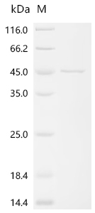
Recombinant Human Syncytin-2 (ERVFRD-1) is produced in E. coli and comprises amino acids 16-350 with a C-terminal 6xHis tag for simplified purification. This partially expressed protein shows purity exceeding 90%, as verified by SDS-PAGE analysis. It's designed for research use only and appears to deliver reliable performance in various experimental applications.

Syncytin-2, encoded by the ERVFRD-1 gene, is an envelope protein derived from endogenous retroviruses. The protein plays what seems to be a crucial role in human placental development. It's integral to syncytiotrophoblast formation, contributing to cell fusion processes. Syncytin-2 has drawn significant interest in studies related to reproductive biology and the evolutionary impact of viral elements in human genomes.

Potential Applications

Note: The applications listed below are based on what we know about this protein's biological functions, published research, and experience from experts in the field. However, we haven't fully tested all of these applications ourselves yet. We'd recommend running some preliminary tests first to make sure they work for your specific research goals.

Based on the provided information, the recombinant human Syncytin-2 is expressed in E. coli, a prokaryotic system that is fundamentally unsuitable for producing functional eukaryotic viral envelope proteins. Syncytin-2 is a complex fusogenic protein that requires precise folding, disulfide bond formation, glycosylation, and correct membrane integration for its biological activity. The protein is expressed as a partial fragment (16-350aa) with a C-terminal 6xHis tag and >90% purity. However, E. coli lacks the eukaryotic chaperones, disulfide isomerases, and glycosylation machinery necessary for proper folding of this retroviral envelope protein. The C-terminal His-tag may further interfere with the transmembrane domain and protein function. Since activity is unverified, the protein cannot be assumed to be correctly folded or bioactive without experimental validation of its fusogenic activity.

1. Protein-Protein Interaction Studies Using His-Tag Pull-Down Assays

The C-terminal 6xHis tag enables technical feasibility for pull-down assays. However, if Syncytin-2 is misfolded (as expected in E. coli), it will not interact physiologically with true binding partners (e.g., ASCT2 receptor). The fusogenic domain requires precise conformation for specific receptor interactions. Identified interactions could be non-physiological artifacts. This application should not be pursued without confirmation of proper folding and receptor-binding capability.

2. Antibody Development and Validation

The recombinant Syncytin-2 can serve as an effective immunogen for generating antibodies that recognize linear epitopes, even if misfolded. The high purity supports immunization protocols. However, antibodies may not recognize conformational or glycosylation-dependent epitopes of native, properly folded Syncytin-2 in human tissues. Validation against endogenous Syncytin-2 is essential.

3. Biochemical Characterization and Stability Studies

This application is well-suited for assessing the recombinant human Syncytin-2 itself. Techniques like circular dichroism spectroscopy, size-exclusion chromatography, and thermal shift assays can evaluate the protein's folding state and stability. These studies are valuable even if the protein is inactive, as they characterize the recombinant product and inform about its suitability for other applications.

4. Comparative Structural Analysis Using Biophysical Methods

This application can provide structural insights, but with significant limitations. Biophysical techniques can assess the E. coli-expressed Syncytin-2's properties, but results will not reflect the native Syncytin-2 structure due to a lack of glycosylation and potential misfolding. The partial sequence (16-350aa) lacks important functional domains, limiting the biological relevance of structural findings.

Final Recommendation & Action Plan
Given the high probability of misfolding in E. coli for this complex viral envelope protein, we recommend first performing comprehensive biophysical characterization (circular dichroism for secondary structure, size-exclusion chromatography for oligomeric state) to assess folding quality. Antibody development can proceed as the safest application. Avoid all functional studies (interactions, fusion assays) until proper folding is validated through receptor-binding or cell-cell fusion assays. For reliable Syncytin-2 research, obtain the protein from mammalian expression systems capable of proper glycosylation and folding. Always include appropriate controls, such as known fusogenic proteins, and validate findings with native Syncytin-2 from human tissues when possible.

Customer Reviews and Q&A

 Customer Reviews

There are currently no reviews for this product.

Submit a Review here

Target Background

Function
This endogenous retroviral envelope protein has retained its original fusogenic properties and participates in trophoblast fusion and the formation of a syncytium during placenta morphogenesis. The interaction with MFSD2A is apparently important for this process.; Endogenous envelope proteins may have kept, lost or modified their original function during evolution but this one can still make pseudotypes with MLV, HIV-1 or SIV-1 virions and confer infectivity. Retroviral envelope proteins mediate receptor recognition and membrane fusion during early infection. The surface protein mediates receptor recognition, while the transmembrane protein anchors the envelope heterodimer to the viral membrane through one transmembrane domain. The other hydrophobic domain, called fusion peptide, mediates fusion of the viral membrane with the target cell membrane.
Gene References into Functions
  1. genetic predisposition in ERVFRDE-1 may be associated with an increased risk of preeclampsia. This polymorphism is possibly involved in the regulation of syncytin-2 expression in preeclamptic placenta. PMID: 29750965
  2. N-glycans at residues 133, 312, 332 and 443 of syncytin-2 are required for optimal fusion induction, and that single-nucleotide polymorphism C46R, N118S, T367M, R417H, V483I and T522M can alter the fusogenic function of syncytin-2. PMID: 26853155
  3. Decreased syncytin-2 and MFSD2 proteins in gestational diabetic placentas might cause abnormal syncytiotrophoblast formation and possibly be involved in the pathology PMID: 26875564
  4. ERVWE1, ERVFRDE1 and ERV3 transcription was down-regulated in hydatidiform moles and gestational trophoblastic neoplasia. PMID: 26992684
  5. These results thereby demonstrate that induced expression of Syncytin-2 is highly dependent on the interaction of bZIP-containing transcription factors to a CRE/AP-1 motif and that this element is important for the regulation of Syncytin-2 expression PMID: 25781974
  6. MFSD2a, the Syncytin-2 receptor, is important for trophoblast fusion. PMID: 23177091
  7. results further highlighted the existence of a correlation between the extent of the decrease in the expression levels of both syncytins 1 and 2 fusogenic proteins and the degree of severity of preeclampsia symptoms PMID: 21493955
  8. Analysis of non-spliced ERVFRDE1 mRNAs and env mRNAs detected efficient splicing of endogenously expressed RNAs in trophoblastic but not in non-placental cells. PMID: 21771862
  9. The crystal structure of a central fragment of syncytin 2 "fossil" ectodomain is reported, allowing a remarkable superposition with the structures of the corresponding domains of present-day infectious retroviruses PMID: 16140326
  10. immunolocalized only in the villous trophoblast of the chorionic villi, at the level of cytotrophoblastic cells PMID: 16714059
  11. findings show that in both humans and mice, one of the two syncytins (human syncytin-2 and mouse syncytin-B) is immunosuppressive and, rather unexpectedly, the other (human syncytin-1 and mouse syncytin-A) is not PMID: 18077339
  12. Syncytin 2 expression illustrates the abnormal trophoblast differentiation observed in placenta of fetal T21-affected pregnancies. PMID: 18215254
  13. expression decreased in preeclamptic placentas; may function as a second fusogenic protein for placental cell fusion PMID: 18650494
  14. These results highlight the importance of Syncytin-2 in BeWo and primary human trophoblast cell fusion. PMID: 19616006

Show More

Hide All

Subcellular Location
Virion.; [Surface protein]: Cell membrane; Peripheral membrane protein.; [Transmembrane protein]: Cell membrane; Single-pass membrane protein.
Protein Families
Gamma type-C retroviral envelope protein family, HERV class-I FRD env subfamily
Tissue Specificity
Expressed at higher level in placenta. Expressed at lower level in adrenal, bone marrow, brain, breast, colon, kidney, lung, ovary, peripheral blood lymphocytes, prostate, skin, spleen, testis, thymus, thyroid, trachea.
Database Links

HGNC: 33823

OMIM: 610524

KEGG: hsa:405754

STRING: 9606.ENSP00000420174

UniGene: Hs.631996

icon of phone
Call us
301-363-4651 (Available 9 a.m. to 5 p.m. CST from Monday to Friday)
icon of address
Address
7505 Fannin St., Ste 610, Room 7 (CUBIO Innovation Center), Houston, TX 77054, USA
icon of social media
Join us with

Subscribe newsletter

Leave a message

* To protect against spam, please pass the CAPTCHA test below.
CAPTCHA verification
© 2007-2025 CUSABIO TECHNOLOGY LLC All rights reserved. 鄂ICP备15011166号-1
×
Place an order now

I. Product details

*
*
*
*

II. Contact details

*
*

III. Ship To

*
*
*
*
*
*
*

IV. Bill To

*
*
*
*
*
*
*
*