Call us
301-363-4651 (Available 9 a.m. to 5 p.m. CST from Monday to Friday)
With the progress of science and medicine, antibodies have become potent tools to recognize, detect, isolate, or visualize their corresponding antigens in basic science research and clinical assays. Whether antibodies work and are stable and reliable in experiments is critical to researchers, clinical users, antibody manufacturers, and journal publishers. It has been reported that ample antibody-based research failed to meet expectations. Therefore, it is necessary to perform functional verification of antibodies to ensure their quality.
This article mainly focuses on antibody validation, which will be elaborated on several aspects, including the definition, significance, methods of antibody validation, and their comparison.
Table of Contents
Antibody validation is the process of confirming whether an antibody can specifically bind to the target antigen through a series of experimental methods or technologies. It should be demonstrated to be specific, selective, and reproducible in the context where the antibody is to be used.
Antibody validation ensures the specificity of the antibody (including the corresponding target antigen, the species of the antigen, etc.), improves the consistency and reproducibility of experimental results, eliminates invalid or unqualified antibodies (save time and cost), confirms the effectiveness and reliability of the antibody in specific applications (such as WB, ELISA, IHC, FC, etc.). Antibody validation should be carried out in the context of specific applications and uses. All applications must be validated independently.
A non-validated antibody may bind to proteins other than their target protein thus producing false positives or may fail to bind to the target protein thus generating false negatives. These problems could lead to invalid experiment data. It is estimated that the life sciences sector loses $800 million a year due to poor antibody quality.
The assessment of antibody validation is based on several factors, including the characterization of antibodies and antigens, binding specificity and selectivity, concentrations of antibodies and additives, documentation, affinity constant, influence of non-target substances, stabilization and storage, application protocols, and user feedback [1].
Antibody validation should minimally include four main criteria: binding specificity, affinity, selectivity, and reproducibility [2].
Specificity: It can be regarded as a measure of the goodness of fit between paratope and epitope, or represents the ability of the antibody to discriminate similar or even dissimilar antigens in the intended application. An antibody with low specificity binds to several different epitopes.
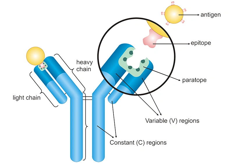
Figure 1: The specific binding between an antibody and an antigen
Affinity: It measures the intensity of the interaction between an antibody and an antigen (epitope). A high-affinity antibody firmly binds to a specific antigen, while a low-affinity antibody weakly binds to the antigen. The higher the affinity of an antibody, the higher the sensitivity of an antibody-based assay.
The equilibrium association constant (Ka) is the basic parameter to evaluate the binding affinity. The Ka is the ratio of antibodies association rate (Kon) to the antibody dissociation rate (Koff). Compared with a low-affinity antibody with a low Ka, a high-affinity antibody with a high Ka will bind more antigens in a shorter period.
Selectivity: It describes how well an antibody binds to its intended target antigen within a complex mixture. An antibody selectivity for a certain antigen means that it shows little cross-reactivity with other antigens.
Reproducibility: It means that the validation data can be reproduced in any lab. However, antibodies vary from batch to batch. The batch-to-batch variability can produce significantly differing results. Compared with polyclonal antibodies and monoclonal antibodies, recombinant antibodies exhibit great superiority in reproducibility.
Generally speaking, what we usually refer to as antibody validation actually refers in most cases to the validation of antibody specificity.
Validation of antibodies needs to meet the applicability of an antibody in a specific application. Frequently used methods include Western blot (WB) antibody validation, ELISA antibody validation, immunofluorescence (IF) antibody validation, immunohistochemistry (IHC) antibody validation, immunocytochemistry (ICC) antibody validation, immunoprecipitation (IP)-mass spectroscopy (MS) antibody validation, flow cytometry (FC) antibody validation, knockout cell line, and knockdown cell line. Here is the list of the comparison among different methods.
Table 1. Antibody validation methods
| Validation types | Detection mechanism | Advantages | Disadvantages |
|---|---|---|---|
| WB | Proteins in a sample are detected through initial molecular weight separation after SDS-PAGE and subsequently blotted on a membrane, finally visualized through a proper antibody (e.g. colorimetric, chemiluminescent, fluorescent, and radioactive detection) |
|
|
| ELISA | Proteins in a sample are detected via specific antibodies and secondary enzyme-conjugated antibody |
|
|
| IMS | Protein complexes are first immunoprecipitated from a cell lysate and then separated via SDS-PAGE, followed by excision of protein bands of interest, and finally analyzed with mass spectrometry |
|
|
| IHC | Proteins in tissue sections are detected via specific antibodies and the antigen-antibody reaction is visualized by color-labeled secondary antibody |
|
|
| ICC | Proteins in single layers of cells grown in culture or from a patient sample are detected via specific antibodies and the antigen-antibody reaction is visualized by fluorophore-labeled secondary antibody |
|
|
| FC | When the target antigen is recognized by the fluorescence-labeled special antibody, the fluorescence signal will be acquired by Flow cytometry |
|
|
| IF | The target protein is detected by a specific fluorescence-labeled antibody, and the fluorescence signal is observed under a fluorescence microscope |
|
|
| Knockout (KO) cell line | Cell lines where the protein-encoding gene of interest is deleted with genetic tools such as CRISPR |
|
|
| Knockdown (KD) cell line | Protein-encoding gene expression is lowered using post-transcriptional gene regulation tools, such as siRNA |
|
|
The International Working Group for Antibody Validation (IWGAV) proposed five conceptual pillars for the validation of antibodies in specific applications [2][4].
(1) Genetic strategies
Genetic strategies employ gene knockout (KO) or gene knockdown (KD) technology to verify the specificity of antibodies. Scientists can use gene editing technologies such as CRISPR/Cas9 or RNAi to knock out or knock down the target gene and then use specific antibodies for detection. If the antibody's binding signal is not observed or significantly reduced in the knocked-out or down cell lines, it indicates that the antibody has good specificity.
Genetic strategies are suitable for antibody specificity validation through WB, IHC, ICC, FS (flow sorting and analysis of cells), ELISA, IP/ChIP (chromatin immunoprecipitation), and RP (reverse-phase protein arrays). However, they can not used for human tissue samples and body fluids such as plasma and serum.
(2) Orthogonal strategies
Orthogonal strategies aim to validate antibody specificity by employing an antibody-independent technique for quantifying the target across multiple samples, followed by analyzing the correlation between this method and antibody-based target quantification. Multiple techniques such as immunoprecipitation (IP), mass spectrometry (MS), and WB can be combined to assess the specificity of the same antibody, providing a more comprehensive validation. Confidence in the specificity of an antibody can be enhanced if multiple methods yield consistent results.
Orthogonal strategies are recommended for antibody validation through WB, IHC, ICC, FS, ELISA, and RP.
(3) Independent antibody strategies
The independent antibody approach uses two or more independent antibodies (antibodies from different sources or lots) to recognize nonoverlapping epitopes on the same protein, and then confirm the specificity by comparison and quantitative analysis.
Independent antibody validation is recommended in the applications, including WB, IHC, ICC, FS, ELISA, IP/ChIP, and RP.
(4) Expression of tagged proteins
This strategy emphasizes the use of verified antibodies as controls to compare the performance of antibodies to be verified. Modify the endogenous target gene, that is, adding the affinity tag (FLAG, V5, etc.) or fluorescent protein (green fluorescent protein) to the endogenous gene, and then detect and compare the signal from the tagged protein using verified antibodies and antibodies to be tested.
This validation strategy can be used in WB, IHC, ICC, and FS applications for antibody specificity validation.
(5) IMS
IP can be used to isolate the target protein by using antibodies to bind specifically to the target protein. Its combination with MS analysis (IMS) can identify target proteins that interact directly with purified antibodies or proteins that may form complexes with target proteins. If the precipitation is successful and the target protein is subsequently detected, the effectiveness of the antibody can be proven. This strategy can be used in IP/CHIP applications.
To ensure the effectiveness and reliability of antibodies, researchers should follow some key practices.
Positive controls are tissue sections or cell lines known to express the target protein. Non-expressing cells that have been transfected with the protein of interest offer the finest positive controls [2]. Positive controls provide a benchmark for experimental results, and researchers can evaluate the reaction strength and specificity of experimental samples by comparing with the results of positive controls.
Negative controls are cell or tissue samples that do not express the target protein. Negative controls can reveal whether there is nonspecific binding or false positive reactions of antibodies. The results of negative controls provide support for the validity of the experiment, ensuring that the observed signal is caused by the specific binding of the antibody to the target antigen and not caused by other factors.
Knockout cells or animals offer the most effective negative controls. VG Magupalli et al. validated anti-NLRP3 and anti-ASC antibodies with respective knockout cells [3]. There are alternative methods that can be utilized to produce comparable control results because these reagents are frequently out of the price range of many labs.
Frequently, it could find cell lines that have been biologically shown to not express a certain protein of interest. For instance, PTEN-null H1650 cells serve as an excellent negative control for PTEN antibodies. siRNA or shRNA knockdown controls are also used as negative controls. When a proper negative control is not available, a cell line or tissue expressing only a small amount of the target proteins can serve as an acceptable substitute.
Positive controls can help confirm the specificity of the antibody and the validity of the experimental setup, while negative controls help identify false positive results, thereby avoiding misinterpretation of experimental data. When performing experiments, proper positive and negative controls can help identify potential experimental errors and background signal interference, thus increasing the confidence of the results.
Selecting the best experimental procedure is an important part of ensuring that the antibody passes the validation process. Use appropriate blocking agents and dilution buffers in your experiment to reduce nonspecific binding. You can optimize your experiment by adjusting conditions such as antibody concentration, incubation time, washing time, and storage and use conditions (e.g. buffer, temperature).
Different antibodies may perform differently in specific experiments (WB, ELISA, IHC, FC, etc.) and need to be verified item by item. Use multiple methods to validate the performance of antibodies as much as possible, such as cross-validation through different experimental techniques to improve the credibility of the results.
The characteristics of each batch of antibodies may be different, so each batch of antibodies should be independently validated. Confirm the stability of antibodies in different production batches to avoid inconsistent experimental results due to batch differences.
Of course, no matter what detection method is used, we need to know in advance the background investigation of the target, including the expression abundance of the target, the spatiotemporal specific expression of the target, the detection threshold of the target antibody, and the appropriate detection method. Only through the analysis of scientific background knowledge, the most suitable method can be selected and a scientific validation strategy can be formulated.
Finally, specific criteria and factors must be met in order to successfully validate and confirm the specificity of an antibody. The specificity of antibodies is dependent on background conditions. Antibodies can only be properly validated in the context of the applied technology and the conditions under which they are ultimately used.
Further reading about knowledge of antibodies:
How to Choose an Antibody for Scientific Research?
How to Choose? Polyclonal, Monoclonal, or Recombinant Antibody?
Cross-reactivity of Antibody: Beneficial or Harmful?
How to Choose the Right Secondary Antibody?
The Overview of Recombinant Antibody
How to Choose the Loading Control Antibodies?
Tag Antibodies, Your Good Partner in Protein Research
Nanobody, what a Powerful Novel Antibody!
How to Choose Monoclonal Antibody Technology? A Definitive Guide
What Are the Best Antibody Purification Methods for Your Research Needs?
References
[1] Michael G Weller. Ten Basic Rules of Antibody Validation [J]. Anal Chem Insights. 2018; 13: 1177390118757462.
[2] Jennifer Bordeaux, Allison W. Welsh, et al. Antibody validation [J]. Biotechniques. 2010 Mar; 48(3): 197–209.
[3] Magupalli V, Negro R, et al. HDAC6 mediates an aggresome-like mechanism for NLRP3 and pyrin inflammasome activation [J]. Science. 2020 Sep 18;369(6510):eaas8995.
[4] Uhlen, M., Bandrowski, A., et al. (2016). A proposal for validation of antibodies [J]. Nature Methods, 13(10), 823.


Comments
Leave a Comment