MLANA Antibody

Code CSB-PA621968ESR1HU
Size US$166
Order now
Image
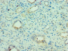
  • Immunohistochemistry of paraffin-embedded human kidney tissue using CSB-PA621968ESR1HU at dilution of 1:100

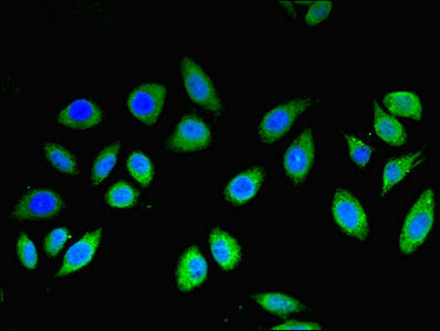
  • Immunofluorescent analysis of A549 cells using CSB-PA621968ESR1HU at dilution of 1:100 and Alexa Fluor 488-congugated AffiniPure Goat Anti-Rabbit IgG(H+L)

Have Questions? Leave a Message or Start an on-line Chat

Product Details

Full Product Name
Rabbit anti-Homo sapiens (Human) MLANA Polyclonal antibody
Uniprot No.
Target Names
MLANA
Alternative Names
Antigen LB39 AA antibody; Antigen LB39-AA antibody; Antigen SK29 AA antibody; Antigen SK29-AA antibody; MAR1_HUMAN antibody; MART 1 antibody; MART-1 antibody; MART1 antibody; Melan A antibody; Melan A protein antibody; Melanoma antigen recognized by T cells 1 antibody; Melanoma antigen recognized by T-cells 1 antibody; MLAN A antibody; MLANA antibody; OTTHUMP00000021036 antibody; OTTHUMP00000021037 antibody; OTTHUMP00000021038 antibody; Protein Melan-A antibody
Raised in
Rabbit
Species Reactivity
Human
Immunogen
Recombinant Human Melanoma antigen recognized by T-cells 1 protein (48-118AA)
Immunogen Species
Homo sapiens (Human)
Conjugate
Non-conjugated
Clonality
Polyclonal
Isotype
IgG
Purification Method
Antigen Affinity Purified
Concentration
It differs from different batches. Please contact us to confirm it.
Buffer
PBS with 0.02% sodium azide, 50% glycerol, pH7.3.
Form
Liquid
Tested Applications
ELISA, IHC, IF
Recommended Dilution
Application Recommended Dilution
IHC 1:20-1:200
IF 1:50-1:200
Troubleshooting and FAQs
Storage
Upon receipt, store at -20°C or -80°C. Avoid repeated freeze.
Lead Time
Basically, we can dispatch the products out in 1-3 working days after receiving your orders. Delivery time maybe differs from different purchasing way or location, please kindly consult your local distributors for specific delivery time.

Customer Reviews and Q&A

 Customer Reviews

There are currently no reviews for this product.

Submit a Review here

Target Background

Function
Involved in melanosome biogenesis by ensuring the stability of GPR143. Plays a vital role in the expression, stability, trafficking, and processing of melanocyte protein PMEL, which is critical to the formation of stage II melanosomes.
Gene References into Functions
  1. Melan-A-stained slides shows definitive invasion. PMID: 27533878
  2. Report automated quantification of proliferation with automated hot-spot selection in phosphohistone H3/MART1 dual-stained stage I/II melanoma. PMID: 27062658
  3. Suggest MelanA-negative spindle-cell associated melanoma constitutes a distinct inflammatory phenotype correlated with dense infiltration of CD163 macrophages and loss of E-cadherin. PMID: 25602697
  4. Inhibin alpha-subunit, Melan A and MNF116 were not sensitive for pheochromocytomas. PMID: 25329118
  5. the presence of antibody and T-cell immune responses directed against the melanocyte-differentiation-antigens MART-1 (Melan-A), tyrosinase and gp100 in patients with melanoma-associated leucoderma, as compared to patients with vitiligo. PMID: 25043574
  6. Knockdown of T-bet expression in Mart-127-35 -specific T-cell-receptor-engineered human CD4(+) CD25(-) and CD8(+) T cells attenuates effector function. PMID: 25495780
  7. Melan-A was useful to confirm the diagnosis of lentigo maligna in early lesions and to differentiate these from chronically sun-damaged skin. PMID: 24394300
  8. Misinitiation of intrathymic MART-1 transcription and biased TCR usage explain the high frequency of MART-1-specific T cells. PMID: 24846220
  9. Comprehensive characterization of the marginal and joint distributions for S100, HMB-45, and Melan-A across a large series of cutaneous melanomas revealed diversity of expression across this group of antigens. PMID: 23899062
  10. melan A (A103) is not expressed in mesotheliomas PMID: 22820661
  11. Report diagnostic role of immunohistochemical staining for Melan-A in evaluating primary melanoma and, specifically, in situ melanoma cases. PMID: 22688397
  12. In this study, production of the C terminus of the Melan-A(26-35) epitope requires cleavage within a hydrophobic stretch, after V35. Hence, the standard proteasome is the only proteasome able to produce the C terminus of this epitope. PMID: 22925930
  13. naturally occurring MART-1 melanoma antigen can be taken-up from dying melanoma cells into DC or MAK and both cell types can induce specific CD8(+) T cell cross-presentation thereafter. PMID: 22768350
  14. The presence of circulating T cells responding to Melan-A or NY-ESO-1 had strong independent prognostic impact on survival in advanced melanoma. PMID: 22529253
  15. The modified melan-A long peptides (not their wild-type counterparts) give rise to highly sustained cross-presentation capacity by monocyte-derived dendritic cells and could be used as tumor vaccines for treatment of melanoma patients. PMID: 22291187
  16. Aberrant expression of Melan-A and MART-1 in AFX and undifferentiated pleomorphic sarcoma of the skin represents an important diagnostic pitfall with potential for misdiagnosis as melanoma. PMID: 22050092
  17. MART-1- and gp100-expressing and -non-expressing melanoma cells are equally proliferative in tumors and clonogenic in vitro. PMID: 21993558
  18. we established several CD4 T-cell clones that recognized a peptide that is embedded within Melan-A/MART-121-50, in a HLA-DPB1*0501 restricted manner PMID: 21760531
  19. Melanocyte MART-1 may correlate to the autoimmune mechanism in children with vitiligo. PMID: 19861278
  20. The differential expression of Mart-1 was investigated in four human uveal melanoma cells. PMID: 21667026
  21. Translocation of the BCL2 gene on the chromosome band 18q21.3 and MYC translocation to 14q32/IGH or less commonly to 2p11/IGK in an identical cell is termed BCL2 and MYC dual-hit lymphoma/leukemia PMID: 21362015
  22. Both semiscale and fullscale quantitative analyses of Ki67/MART1 stains are valuable diagnostic tools to distinguish melanomas and nevi with a large degree of certainty. PMID: 21610457
  23. Molecular upstaging of MART-1 using rt-pcr was correlated with prognosis in melanoma patients PMID: 21135695
  24. determined the crystal structure of a phosphopeptide derived from melanoma antigen recognized by T cells-1 (pMART-1), selectively expressed by melanomas, in complex with HLA-DR1 PMID: 20417641
  25. OA1 interacts with MART-1 at early stages of melanogenesis to control melanosome identity and composition. PMID: 19717472
  26. unexpected high frequency of T cells specific for the self-antigen Melan-A/MART-1 in CD8 single-positive thymocytes from human histocompatibility leukocyte antigen-A2 healthy individuals PMID: 11854361
  27. Melanosomes from an Achilles tendon clear-cell sarcoma showed expression of the Melan-A gene. PMID: 12072203
  28. The high frequency of Melan-A multimer(+) T cells can be explained by the existence of largely cross-reactive subsets of naive CD8(+) T cells displaying multiple specificities. PMID: 12119345
  29. dominant function of TCRAV2 segment in forming the TCR repertoire specific for the human self Ag Melan-A/MART-1 PMID: 12444131
  30. Data show that MITF appears to regulate the expression of the SILV and MLANA genes. PMID: 12819038
  31. Antigen-specific T lymphocyte reactivity to MelanA/MART-1 and tyrosinase peptides was not observed ex vivo in our vitiligo patients, and only one patient demonstrated responses to MelanA/MART-1 PMID: 12925214
  32. adrenocortical neoplasms showed strong and diffuse granular cytoplasmic staining for Melan A. No nuclear reaction was observed. PMID: 14663359
  33. the expression of Melan-A/MART-1 in patient-derived cell cultures may help to identify a group of melanoma patients with prolonged survival PMID: 15305155
  34. Melan-A is very useful in distinguishing between desmoplastic melanoma and sclerotic nevi. PMID: 15618925
  35. Data suggest that MART-1 is indispensable for Pmel17 function and thus plays an important role in regulating mammalian pigmentation. PMID: 15695812
  36. Two new Melan-A-derived CD4+ T cell epitopes have been identified that map to the 1-20 and 91-110 regions of the protein and are restricted by HLA-DR11 and HLA-DR52 molecules. PMID: 16814609
  37. Blocking MAPK signaling augments antigen levels, thereby enhancing Melan-A/MART-1-specific cytotoxic T-cell responses to antigen-negative cells following MEK inhibition treatment. PMID: 17050671
  38. Two novel Melan-A/MART-1-derived sequences recognized by CD4 T cells from melanoma patients provide the basis for monitoring of naturally occurring and vaccine-induced Melan-A/MART-1-specific CD4 T cell responses. PMID: 17082590
  39. HMB-45 and Melan-A combined were positive in 100% of the renal angiomyolipomas. PMID: 17227112
  40. Data show that the MAT-1 26-35 and 27-35 peptides adopt strikingly different conformations when bound to HLA-A2. PMID: 17719062
  41. t cell recaptor antigens and the major histocompatibilyt complex are complexed with this protein. PMID: 17768347
  42. the combinational characterization of calretinin (either by polyclonal or monoclonal antibody), inhibin alpha, and Melan-A expression is of great significance in the differential diagnosis of adrenocortical tumors. PMID: 18091323
  43. these data significantly enlarge the fraction of melanoma patients susceptible to benefit from a Melan-A/MART1 vaccine approach. PMID: 18097665
  44. Measurement of Melan-A, gp100, MAGE-3, MIA and tyrosinase represents a prognostic factor and a method for early detection of metastasis and treatment response of melanoma patients. PMID: 18181974
  45. Although melan-A-specific T cells were detectable, they are unlikely to be involved in the etiopathogenesis of vitiligo. PMID: 18337837
  46. Increased staining with Melan-A, in an actinic keratosis, or solar lentigo should raise the possibility of a contiguous melanoma in situ. PMID: 18494818
  47. results show a complex pattern of changes in the expression of Melan-A in melanomas depending on the location of melanoma cells within individual skin layers PMID: 18626310
  48. the generation of larger numbers of ELAGIGILTV ((MART-1 26-35, A27L)-specific CD8+ T cells correlates negatively with the acquisition of a CD45RA+CCR7- phenotype and cytotoxic capacity. PMID: 19050104
  49. T-cell receptor beta chain sequences of Melan-A-specific clonotypes obtained from melanoma patients were highly heterogeneous, but displayed a preferential usage of few TRBV and TRBJ segments. PMID: 19317896
  50. Case Report: Melan-a-positive "pseudomelanocytic nests": a pitfall in the histopathologic and immunohistochemical diagnosis of pigmented lesions on sun-damaged skin. PMID: 19384076

Show More

Hide All

Subcellular Location
Endoplasmic reticulum membrane; Single-pass type III membrane protein. Golgi apparatus. Golgi apparatus, trans-Golgi network membrane. Melanosome. Note=Also found in small vesicles and tubules dispersed over the entire cytoplasm. A small fraction of the protein is inserted into the membrane in an inverted orientation. Inversion of membrane topology results in the relocalization of the protein from a predominant Golgi/post-Golgi area to the endoplasmic reticulum. Melanoma cells expressing the protein with an inverted membrane topology are more effectively recognized by specific cytolytic T-lymphocytes than those expressing the protein in its native membrane orientation.
Tissue Specificity
Expression is restricted to melanoma and melanocyte cell lines and retina.
Database Links

HGNC: 7124

OMIM: 605513

KEGG: hsa:2315

STRING: 9606.ENSP00000370880

UniGene: Hs.154069

icon of phone
Call us
301-363-4651 (Available 9 a.m. to 5 p.m. CST from Monday to Friday)
icon of address
Address
7505 Fannin St., Ste 610, Room 7 (CUBIO Innovation Center), Houston, TX 77054, USA
icon of social media
Join us with

Subscribe newsletter

Leave a message

* To protect against spam, please pass the CAPTCHA test below.
CAPTCHA verification
© 2007-2024 CUSABIO TECHNOLOGY LLC All rights reserved. 鄂ICP备15011166号-1
webinars: DT3C facilitates antibody internalization X
Place an order now

I. Product details

*
*
*
*

II. Contact details

*
*

III. Ship To

*
*
*
*
*
*
*

IV. Bill To

*
*
*
*
*
*
*
*