Call us
301-363-4651 (Available 9 a.m. to 5 p.m. CST from Monday to Friday)
| Code | CSB-EP3402GND |
| Abbreviation | Recombinant SARS-CoV-2 Envelope small membrane protein&Membrane protein, partial |
| MSDS | |
| Size | $224 |
| Order now | |
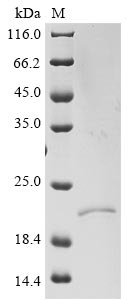
| Image |
|
| Have Questions? | Leave a Message or Start an on-line Chat |
Recombinant Severe acute respiratory syndrome coronavirus 2 Envelope small membrane protein & Membrane protein (E & M) is expressed in E. coli with an N-terminal 6xHis-SUMO tag. This partial protein covers the expression region from 1-42 amino acids and achieves a purity level of over 90% as verified by SDS-PAGE. This product is intended for research use only and does not contain endotoxin levels suitable for clinical applications.
The Envelope (E) and Membrane (M) proteins of Severe acute respiratory syndrome coronavirus 2 appear to play crucial roles in the virus's structure and life cycle. The E protein seems to be involved in virus assembly and release, while the M protein is likely integral to maintaining the virus's shape and size. Both proteins may prove essential for studying viral pathogenesis and developing potential therapeutic interventions.
Potential Applications
Note: The applications listed below are based on what we know about this protein's biological functions, published research, and experience from experts in the field. However, we haven't fully tested all of these applications ourselves yet. We'd recommend running some preliminary tests first to make sure they work for your specific research goals.
The protein is expressed in E. coli as a partial fragment (1–389 aa) of SARS-CoV-2 envelope (E) and membrane (M) proteins, tagged with an N-terminal 6xHis-SUMO motif. While the SUMO tag enhances solubility, E. coli struggles to correctly fold eukaryotic membrane proteins (E and M are integral membrane proteins with complex topology). No data confirms native secondary/tertiary structure (e.g., circular dichroism, thermal shift assays). E. coli may misfold the fragment, leading to aggregation or non-functional conformations. The fragment lacks full-length context (e.g., transmembrane domains, interaction motifs for other viral proteins like Spike [S] or host factors). Bioactivity (e.g., membrane association, protein-protein interactions) is untested and unlikely to mimic native E/M function.
1. Antibody Development and Screening
This recombinant E&M fragment can serve as an immunogen for generating antibodies, but antibody specificity must be validated against native E/M—E. coli-expressed protein may present non-native epitopes, leading to cross-reactivity with irrelevant targets. The His-SUMO tag simplifies purification/immobilization for ELISA, but antibodies may not recognize the fragment in its native context (e.g., on viral particles).
2. Protein-Protein Interaction Studies
Pull-down assays using the His-SUMO tag can identify interactors, but results depend on correct folding—E. coli-expressed fragments often misfold, causing false positives/negatives. Identified partners must be validated via co-IP or functional assays to rule out artifacts. The partial length restricts insights to interactions within this domain, not full-length E/M biology.
3. Structural and Biochemical Characterization
This fragment supports preliminary biophysical studies (e.g., CD for secondary structure, DLS for stability) but cannot inform native E/M architecture—E. coli-expressed protein may lack correct disulfide bonds or transmembrane folding. Structural conclusions (e.g., oligomerization) must be contextualized by folding limitations.
4. ELISA-Based Binding Assays
The His-SUMO tag enables immobilization for ELISA, but misfolding may alter binding—results are qualitative at best. Quantitative affinity measurements require a confirmed native structure, as the fragment’s conformation may not support native-like interactions.
Final Recommendation & Action Plan
This E. coli-expressed SARS-CoV-2 E&M fragment has limited utility without rigorous validation: first, confirm folding via CD spectroscopy and thermal shift assays to rule out misfolding; second, test bioactivity (e.g., membrane association or interaction with Spike) to ensure native-like function. Optimize expression (e.g., co-express chaperones, use low-temperature induction) to improve solubility. For antibody development, validate specificity against native E/M; for interactions/ELISA, use tag cleavage or orthogonal methods (e.g., SPR) to reduce artifacts. If folding/bioactivity fails, use a eukaryotic system (e.g., mammalian/insect cells) to ensure native structure—this fragment alone is insufficient for reliable downstream applications without validation.
There are currently no reviews for this product.

