Call us
301-363-4651 (Available 9 a.m. to 5 p.m. CST from Monday to Friday)
| Code | CSB-MP886444MO |
| Abbreviation | Recombinant Mouse Hebp1 protein |
| MSDS | |
| Size | $9.9 |
| Promotion |

|
| Order now | |
| Image |
|
| Have Questions? | Leave a Message or Start an on-line Chat |
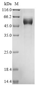
Recombinant Mouse Heme-binding protein 1 (Hebp1) comes from a mammalian cell expression system, which appears to support proper protein folding and post-translational modifications. The full-length protein spans amino acids 1 to 190 and includes a C-terminal hFc1 tag that helps with purification and detection. Purity levels reach above 85%, confirmed through SDS-PAGE analysis. This product is strictly for research purposes and cannot be used in clinical settings.
Heme-binding protein 1 (Hebp1) likely plays an important role in how cells handle heme metabolism. Scientists study this protein to understand its involvement in heme transport and regulation—processes that seem essential for keeping cells balanced and functioning properly. Learning how Hebp1 works may shed light on broader biochemical pathways related to heme use and could have implications across various biological contexts.
Potential Applications
Note: The applications listed below are based on what we know about this protein's biological functions, published research, and experience from experts in the field. However, we haven't fully tested all of these applications ourselves yet. We'd recommend running some preliminary tests first to make sure they work for your specific research goals.
Mouse Hebp1 is a heme-binding protein that requires precise folding and proper heme-binding domain formation for its functional activity in heme homeostasis. The mammalian cell expression system provides a eukaryotic environment that supports proper protein folding, post-translational modifications, and complex assembly, significantly increasing the probability of correct folding compared to prokaryotic systems. However, the C-terminal hFc1 tag (∼25 kDa) is large and may sterically interfere with the protein's functional domains or oligomerization interfaces. While mammalian expression provides favorable conditions, experimental validation remains essential to confirm both structural integrity and functional heme-binding activity.
1. Antibody Development and Validation Studies
Antibody development benefits from correct folding but can proceed based on sequence alone. Antibody development relies on antigenic sequence recognition. If correctly folded (verified), the protein excels for generating conformation-sensitive antibodies that recognize native Hebp1 epitopes. If misfolded/unverified, it remains suitable for producing antibodies against linear epitopes, which are still valuable for detection applications.
2. Protein-Protein Interaction Studies
This application requires proper folding validation. Protein-protein interactions within heme homeostasis pathways require precise tertiary structure. If correctly folded (verified), the protein is suitable for identifying physiological interaction partners. If misfolded/unverified, there is a high risk of non-specific binding or interaction failure, making results biologically misleading.
3. Biochemical Characterization and Binding Assays
These studies are essential for determining folding status and functional competence. Functional heme-binding requires native protein conformation that must be validated. If correctly folded and heme-binding competent (verified), characterization provides reliable data on binding kinetics, specificity, and stoichiometry. If misfolded/inactive (unverified), binding assays will yield negative or misleading results.
4. ELISA-Based Detection and Quantification Assays
ELISA development relies on consistent antigen availability and detection, not functional conformation. This application is well-suited regardless of folding status. Immunoassays depend on antibody-epitope binding rather than functional conformation. If correctly folded (verified), the protein provides authentic antigenic presentation. If misfolded/unverified, it remains effective as a standardized antigen for assay development.
Final Recommendation & Action Plan
The mammalian expression system provides favorable eukaryotic folding conditions for this heme-binding protein, but experimental validation of structural integrity and heme-binding activity is essential before reliable use in functional studies. Begin with Application 3 (Biochemical Characterization) to assess folding quality through size-exclusion chromatography, circular dichroism spectroscopy, and validate heme-binding activity using spectroscopic methods. Once correct folding and functional activity are verified, proceed cautiously with Applications 1, 2, and 4 for antibody development, interaction studies, and assay development. If misfolding or lack of heme-binding is detected, limit applications to linear epitope antibody production (Application 1) and ELISA standardization (Application 4), avoiding all functional interaction and binding studies. For reliable Hebp1 research, always include appropriate heme-binding controls and consider the potential steric effects of the large Fc tag in interaction studies.
There are currently no reviews for this product.

