Recombinant Mouse Heme-binding protein 1 (Hebp1)

In Stock
Code CSB-EP886444MOb1
Abbreviation Recombinant Mouse Hebp1 protein
MSDS
Size $388
Order now
Image
  • (Tris-Glycine gel) Discontinuous SDS-PAGE (reduced) with 5% enrichment gel and 15% separation gel.
Have Questions? Leave a Message or Start an on-line Chat

Product Details

Purity
Greater than 90% as determined by SDS-PAGE.
Target Names
Hebp1
Uniprot No.
Research Area
Cardiovascular
Alternative Names
(p22HBP)
Species
Mus musculus (Mouse)
Source
E.coli
Expression Region
1-190aa
Target Protein Sequence
MLGMIRNSLFGSVETWPWQVLSTGGKEDVSYEERACEGGKFATVEVTDKPVDEALREAMPKIMKYVGGTNDKGVGMGMTVPVSFALFPNEDGSLQKKLKVWFRIPNQFQGSPPAPSDESVKIEEREGITVYSTQFGGYAKEADYVAHATQLRTTLEGTPATYQGDVYYCAGYDPPMKPYGRRNEVWLVKA
Note: The complete sequence may include tag sequence, target protein sequence, linker sequence and extra sequence that is translated with the protein sequence for the purpose(s) of secretion, stability, solubility, etc.
If the exact amino acid sequence of this recombinant protein is critical to your application, please explicitly request the full and complete sequence of this protein before ordering.
Mol. Weight
28.5 kDa
Protein Length
Full Length
Tag Info
N-terminal 10xHis-tagged and C-terminal Myc-tagged
Form
Liquid or Lyophilized powder
Note: We will preferentially ship the format that we have in stock, however, if you have any special requirement for the format, please remark your requirement when placing the order, we will prepare according to your demand.
Buffer
If the delivery form is liquid, the default storage buffer is Tris/PBS-based buffer, 5%-50% glycerol. If the delivery form is lyophilized powder, the buffer before lyophilization is Tris/PBS-based buffer, 6% Trehalose.
Reconstitution
We recommend that this vial be briefly centrifuged prior to opening to bring the contents to the bottom. Please reconstitute protein in deionized sterile water to a concentration of 0.1-1.0 mg/mL.We recommend to add 5-50% of glycerol (final concentration) and aliquot for long-term storage at -20°C/-80°C. Our default final concentration of glycerol is 50%. Customers could use it as reference.
Troubleshooting and FAQs
Storage Condition
Store at -20°C/-80°C upon receipt, aliquoting is necessary for mutiple use. Avoid repeated freeze-thaw cycles.
Shelf Life
The shelf life is related to many factors, storage state, buffer ingredients, storage temperature and the stability of the protein itself.
Generally, the shelf life of liquid form is 6 months at -20°C/-80°C. The shelf life of lyophilized form is 12 months at -20°C/-80°C.
Lead Time
3-7 business days
Notes
Repeated freezing and thawing is not recommended. Store working aliquots at 4°C for up to one week.
Datasheet & COA
Please contact us to get it.
Description

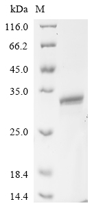
Recombinant Mouse Heme-binding protein 1 (Hebp1) comes from an E.coli expression system and spans the complete protein sequence from amino acids 1 to 190. The construct includes an N-terminal 10xHis tag and a C-terminal Myc tag, which makes purification and detection more straightforward. SDS-PAGE analysis confirms the product achieves greater than 90% purity - a level that appears suitable for most research needs.

Heme-binding protein 1 (Hebp1) binds heme, though the full scope of this interaction remains an active area of investigation. This protein likely contributes to heme metabolism and may play a part in how cells respond to oxidative stress. Scientists are working to better understand Hebp1's role in these pathways, along with its broader involvement in heme-related cellular processes.

Potential Applications

Note: The applications listed below are based on what we know about this protein's biological functions, published research, and experience from experts in the field. However, we haven't fully tested all of these applications ourselves yet. We'd recommend running some preliminary tests first to make sure they work for your specific research goals.

Mouse Hebp1 is a eukaryotic heme-binding protein that requires precise folding and proper heme-binding domain formation for its functional activity. The E. coli expression system cannot provide the necessary eukaryotic folding environment, post-translational modifications, and molecular chaperones required for this protein. While the protein may be soluble, the dual N-terminal 10xHis-tag and C-terminal Myc-tag may sterically interfere with the protein's functional domains. 

1. Antibody Development and Validation Studies

This application is highly suitable as antibody development relies on antigenic sequence recognition rather than functional folding. If correctly folded (verified), the protein excels for generating conformation-sensitive antibodies. If misfolded/unverified, it remains highly suitable for producing antibodies against linear epitopes.

2. Tag-Assisted Purification and Detection Assays

This application is well-suited regardless of folding status. Tag-based systems rely on tag functionality rather than native protein folding. The dual tags provide excellent flexibility for purification and detection method development.

3. Biochemical Characterization and Stability Studies

These studies are essential for determining folding status. If correctly folded (verified), characterization provides reliable data on structural properties. If misfolded/unverified, analysis yields physical property data for quality control.

Final Recommendation & Action Plan

The E. coli expression system is fundamentally limited for this eukaryotic protein due to folding environment differences. Begin with Application 3 (Biochemical Characterization) to assess folding quality through techniques like circular dichroism spectroscopy and size-exclusion chromatography. Applications 1 and 2 (antibody development and tag-based assays) can proceed immediately as they don't require native folding. Protein-protein interactions require a precise tertiary structure that E. coli may not provide. If misfolding is detected, limit applications to linear epitope antibody production and tag-based method development.

Customer Reviews and Q&A

 Customer Reviews

There are currently no reviews for this product.

Submit a Review here

Target Background

Function
May bind free porphyrinogens that may be present in the cell and thus facilitate removal of these potentially toxic compound. Binds with a high affinity to one molecule of heme or porphyrins. It binds metalloporphyrins, free porphyrins and N-methylprotoporphyrin with similar affinities.
Gene References into Functions
  1. Results present a systematic molecular modeling study of the binding properties of murine and human p22HBP protein (heme-binding protein) with four tetrapyrrole ring systems belonging to the heme biosynthetic pathway. PMID: 20800521
  2. Characterization as a tetrapyrrole-binding protein PMID: 12413491
  3. The heme-binding properties and coordination structure of mouse liver p22HBP are distinct from those of SOUL protein, despite high sequence homology. PMID: 15518569
  4. analysis of murine P22HBP structure PMID: 16905545
Subcellular Location
Cytoplasm.
Protein Families
HEBP family
Tissue Specificity
Ubiquitously expressed. Extremely abundant in liver.
Database Links
icon of phone
Call us
301-363-4651 (Available 9 a.m. to 5 p.m. CST from Monday to Friday)
icon of address
Address
7505 Fannin St., Ste 610, Room 7 (CUBIO Innovation Center), Houston, TX 77054, USA
icon of social media
Join us with

Subscribe newsletter

Leave a message

* To protect against spam, please pass the CAPTCHA test below.
CAPTCHA verification
© 2007-2025 CUSABIO TECHNOLOGY LLC All rights reserved. 鄂ICP备15011166号-1
×
Place an order now

I. Product details

*
*
*
*

II. Contact details

*
*

III. Ship To

*
*
*
*
*
*
*

IV. Bill To

*
*
*
*
*
*
*
*