Call us
301-363-4651 (Available 9 a.m. to 5 p.m. CST from Monday to Friday)
| Code | CSB-RA272392A0HU |
| Size | US$210 |
| Order now | |
| Image |
|
| Have Questions? | Leave a Message or Start an on-line Chat |
| Application | Recommended Dilution |
|---|---|
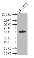
| WB | 1:500-1:2000 |
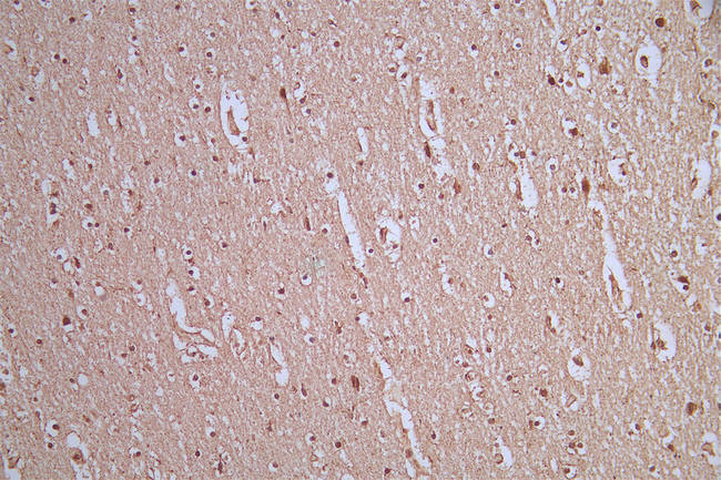
| IHC | 1:50-1:200 |
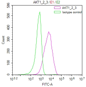
| FC | 1:50-1:200 |
The AKT1/2/3 recombinant monoclonal antibody is created using in vitro expression systems, which are established by cloning the DNA sequences of AKT1/2/3 antibodies obtained from immunoreactive rabbits. The immunogen used in this process is a synthesized peptide derived from the human AKT1/2/3 protein. Subsequently, the genes encoding the AKT1/2/3 antibodies are inserted into plasmid vectors, and these recombinant plasmid vectors are transfected into host cells to facilitate antibody expression. The AKT1/2/3 recombinant monoclonal antibody then undergoes affinity-chromatography purification and is rigorously tested for functionality in ELISA, WB, IHC, and FC applications, confirming its reactivity with the human AKT1/2/3 protein.
AKT1, AKT2, and AKT3 are closely related isoforms of the AKT protein kinase family. While they share structural similarities and some functional overlap, they exhibit distinct tissue-specific expression patterns and play specialized roles in various cellular processes. AKT1 is widely expressed in various tissues and is involved in the regulation of cell growth, survival, and proliferation. AKT2 is predominantly expressed in insulin-responsive tissues such as skeletal muscle, liver, and adipose tissue, and plays a crucial role in glucose homeostasis and insulin signaling. AKT3 is expressed at higher levels in the brain and nervous system, where it contributes to neuronal development and function.
There are currently no reviews for this product.

