Call us
301-363-4651 (Available 9 a.m. to 5 p.m. CST from Monday to Friday)
| Code | CSB-RA159926A0HU |
| Size | US$210 |
| Order now | |
| Image |
|
| Have Questions? | Leave a Message or Start an on-line Chat |
| Application | Recommended Dilution |
|---|---|
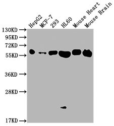
| WB | 1:500-1:5000 |
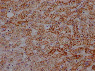
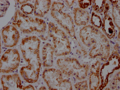
| IHC | 1:50-1:200 |
ATP5A1, also known as ATP synthase subunit alpha, serves as a critical component of the mitochondrial F1F0-ATP synthase complex responsible for the majority of cellular ATP production. This enzyme complex plays a fundamental role in oxidative phosphorylation, making ATP5A1 an essential marker for studying mitochondrial function, cellular metabolism, and bioenergetics. Dysregulation of ATP synthase activity has been implicated in various pathological conditions, including cancer metabolism alterations and neurodegenerative diseases, positioning this target at the intersection of multiple active research fields.
This recombinant monoclonal antibody, generated from clone 5G11, offers the reproducibility and consistency that demanding experimental workflows require. Because recombinant antibodies are produced from defined sequences rather than traditional hybridoma methods, researchers benefit from lot-to-lot uniformity that supports longitudinal studies and ensures comparable results across experiments.
Validation studies demonstrate robust performance across multiple applications. In western blot analyses at 1:2000 dilution, this antibody detects a clean 60 kDa band consistent with the predicted molecular weight of ATP5A1. Detection has been confirmed in diverse human cell lines including HepG2, MCF-7, 293, and HL60, providing flexibility for researchers working with different cellular models. Cross-species reactivity extends to mouse tissues, with successful detection demonstrated in both heart and brain lysates. For immunohistochemistry applications, validation in paraffin-embedded human liver and kidney tissues at 1:100 dilution shows reliable staining using standard citrate buffer antigen retrieval protocols.
Whether investigating mitochondrial dysfunction in cancer models, exploring metabolic signaling pathways, or characterizing tissue-specific expression patterns, this antibody provides a dependable tool for ATP5A1 detection across your experimental platforms.
There are currently no reviews for this product.

