Call us
301-363-4651 (Available 9 a.m. to 5 p.m. CST from Monday to Friday)
| Code | CSB-RA556985A0HU |
| Size | US$210 |
| Order now | |
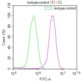
| Image |
|
| Have Questions? | Leave a Message or Start an on-line Chat |
| Application | Recommended Dilution |
|---|---|
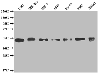
| WB | 1:500-1:2000 |
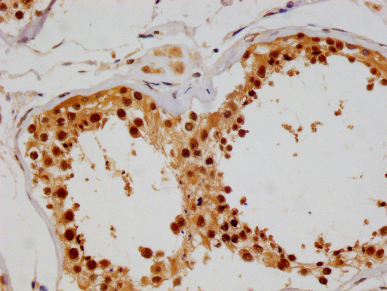
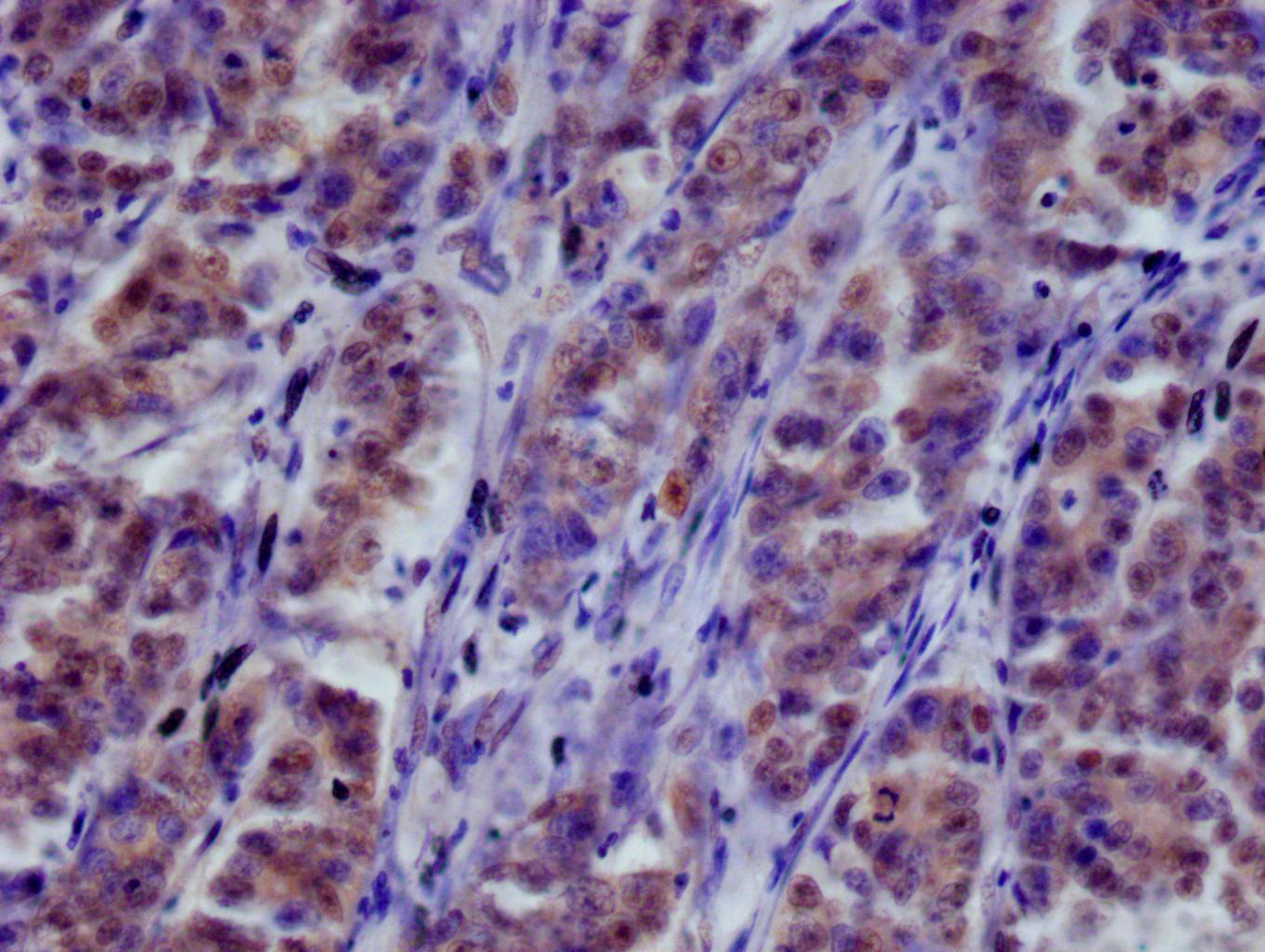
| IHC | 1:50-1:200 |
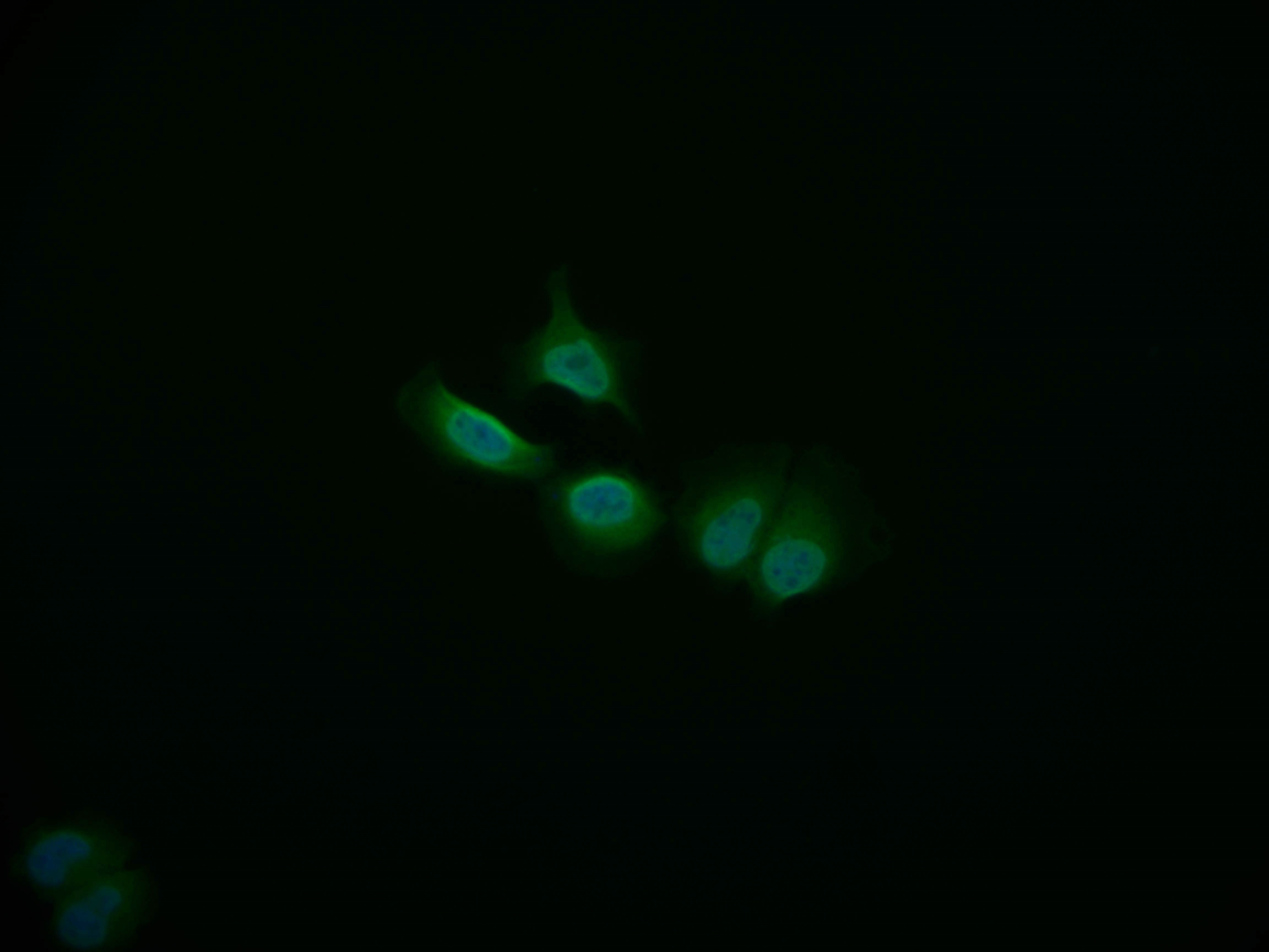
| IF | 1:50-1:200 |
| FC | 1:50-1:200 |
To produce the COPS3 recombinant monoclonal antibody, a series of complex processes is required. First, the COPS3 monoclonal antibody is collected and its gene sequence is determined. A COPS3 monoclonal antibody gene-carrying vector is then constructed and introduced into a host cell line for culture. A synthesized peptide from human COPS3 is used as an immunogen during COPS3 monoclonal antibody production. Affinity chromatography is employed to purify the resulting COPS3 recombinant monoclonal antibody, which is then evaluated for specificity using multiple applications, including ELISA, WB, IHC, IF, and FC.
The COPS3 recombinant monoclonal antibody can react with human COPS3 protein.COPS3, also known as CSN3, is a component of the COP9 signalosome complex (CSN) which is a highly conserved protein complex found in eukaryotic cells. The CSN is involved in the regulation of various cellular processes such as protein degradation, cell cycle control, DNA repair, and signal transduction. COPS3 plays a role in the stability and activity of the CSN complex, and its function is essential for the proper function of the complex.
There are currently no reviews for this product.

