DCP1A Recombinant Monoclonal Antibody

Code CSB-RA109371A0HU
Size US$210
Order now
Image
  • Western Blot
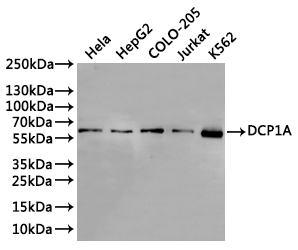
    Positive WB detected in: Hela whole cell lysate(30µg), HepG2 whole cell lysate(30µg), COLO-205 whole cell lysate(30µg), Jurkat whole cell lysate(30µg), k562 whole cell lysate(30µg)
    All lanes: DCP1A antibody at 1:1000
    Secondary
    Goat polyclonal to rabbit IgG at 1/40000 dilution
    Predicted band size: 63 kDa
    Observed band size: 63 kDa
    Exposure time:2min
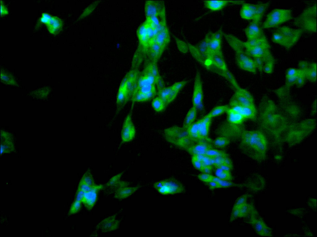
  • Immunofluorescence staining of SH-SY5Y cell with CSB-RA109371A0HU at 1:50, counter-stained with DAPI. The cells were fixed in 4% formaldehyde, permeabilized using 0.2% Triton X-100 and blocked in 10% normal Goat Serum. The cells were then incubated with the antibody overnight at 4°C. The secondary antibody was Alexa Fluor 488-congugated AffiniPure Goat Anti-Rabbit IgG(H+L).
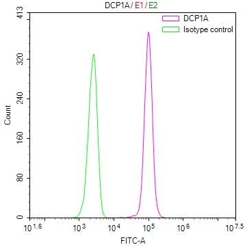
  • Overlay Peak curve showing jurkat cells stained with CSB-RA109371A0HU (red line) at 1:100. The cells were fixed in 4% formaldehyde and permeated by 0.2% TritonX-100 for 10min. Then 10% normal goat serum to block non-specific protein-protein interactions followed by the antibody (1ug/1*106cells) for 45min at 4℃. The secondary antibody used was FITC-conjugated goat anti-rabbit IgG (H+L) at 1/200 dilution for 35min at 4℃.Control antibody (green line) was Rabbit IgG (1ug/1*106cells) used under the same conditions. Acquisition of >10,000 events was performed.
Have Questions? Leave a Message or Start an on-line Chat

Product Details

Uniprot No.
Target Names
DCP1A
Alternative Names
mRNA-decapping enzyme 1A (EC 3.-.-.-) (Smad4-interacting transcriptional co-activator) (Transcription factor SMIF), DCP1A, SMIF
Species Reactivity
Human
Immunogen
Recombinant Antibody
Immunogen Species
Homo sapiens (Human)
Conjugate
Non-conjugated
Clonality
Monoclonal
Isotype
Rabbit IgG
Clone No.
10E12
Purification Method
Affinity-chromatography
Concentration
It differs from different batches. Please contact us to confirm it.
Buffer
Rabbit IgG in 10mM phosphate buffered saline , pH 7.4, 150mM sodium chloride, 0.05% BSA, 0.02% sodium azide and 50% glycerol.
Form
Liquid
Tested Applications
ELISA, WB, IF, FC
Recommended Dilution
Application Recommended Dilution
WB 1:500-1:5000
IF 1:50-1:200
FC 1:50-1:200
Troubleshooting and FAQs
Storage
Upon receipt, store at -20°C or -80°C. Avoid repeated freeze.
Lead Time
Basically, we can dispatch the products out in 1-3 working days after receiving your orders. Delivery time maybe differs from different purchasing way or location, please kindly consult your local distributors for specific delivery time.
Description

DCP1A, also known as mRNA-decapping enzyme 1A, plays a central role in mRNA turnover by catalyzing the removal of the 5' cap structure, a critical step that commits transcripts to degradation. Beyond its function in mRNA decay, DCP1A also serves as a Smad4-interacting transcriptional co-activator (SMIF), linking it to TGF-β signaling pathways. This dual functionality makes DCP1A a compelling target for researchers investigating post-transcriptional gene regulation, stress granule dynamics, and cellular signaling networks.

This recombinant monoclonal antibody, generated in rabbit using clone 10E12, offers the reproducibility and consistency that demanding experimental workflows require. Because recombinant antibodies are produced from defined sequences, you can expect uniform performance across lots, eliminating the variability often encountered with traditional hybridoma-derived reagents. The antibody has been affinity-purified to ensure high specificity for its target.

Validation studies demonstrate robust performance across multiple applications. Western blot analysis detects a band at the predicted molecular weight of 63 kDa across diverse human cell lines including HeLa, HepG2, COLO-205, Jurkat, and K562, confirming reliable detection in both epithelial and hematopoietic cell contexts. For immunofluorescence microscopy, the antibody successfully visualizes DCP1A localization in SH-SY5Y neuroblastoma cells at dilutions of 1:50 to 1:200. Flow cytometry validation in Jurkat cells shows clear separation between specific signal and isotype control, enabling quantitative analysis of DCP1A expression at the single-cell level.

This antibody is well-suited for studies examining P-body formation, mRNA surveillance mechanisms, and the intersection of transcriptional regulation with mRNA decay pathways in human cell systems.

Usage
For Research Use Only. Not for use in diagnostic or therapeutic procedures.

Customer Reviews and Q&A

 Customer Reviews

There are currently no reviews for this product.

Submit a Review here

Target Background

Function
Necessary for the degradation of mRNAs, both in normal mRNA turnover and in nonsense-mediated mRNA decay. Removes the 7-methyl guanine cap structure from mRNA molecules, yielding a 5'-phosphorylated mRNA fragment and 7m-GDP. Contributes to the transactivation of target genes after stimulation by TGFB1. Essential for embryonic development.
Gene References into Functions
  1. Overexpression of DCP1A is associated with malignant melanoma. PMID: 29076924
  2. The results suggest that overexpressed Dcp1a and GW182 can form different cytoplasmic aggregates and play distinct biological roles in the miRNA pathway. PMID: 28488892
  3. The E3 ligase TRAF6 binds to DCP1a and indirectly regulates DCP1a phosphorylation, expression of decapping factors, and gene-specific mRNA decay. PMID: 27315556
  4. DCP1A rs11551405 may have a prognostic effect on survival of CM patients. PMID: 27578485
  5. The assembly of EDC4 and Dcp1a into processing bodies is critical for the translational regulation of IL-6. PMID: 25970328
  6. Phosphorylation at serine 315, serine 319, and threonine 321 of DCP1A modulates IL-8 expression during respiratory syncytial virus infection. PMID: 25796077
  7. The data indicates that DCP2 activation by DCP1 occurs preferentially on the EDC4 scaffold, which may serve to couple DCP2 activation by DCP1 with 5'-to-3' mRNA degradation by XRN1 in human cells. PMID: 24510189
  8. Malin regulates the recruitment of mRNA-decapping enzyme 1A (Dcp1a) to processing bodies. PMID: 23131811
  9. hDcp1a has a role in control of processing body dynamics during the cell cycle via phosphorylation PMID: 23300942
  10. Reveal DCP1a as a multifunctional regulator of mRNA expression and suggest a role for JNK kinase phosphorylation in controlling the subcellular localization of DCP1a in response to stress or inflammatory stimuli. PMID: 21859862
  11. Data show the presence of P-body-like foci in mouse oocytes, as revealed by the presence of Dcp1a and the colocalization of RNA-associated protein 55 (RAP55) and the DEAD box RNA helicase Rck/p54. PMID: 19812249
  12. Overexpression of wild-type SMIF enhanced expression of TGFbeta/BMP regulated genes, whereas a dominant-negative SMIF mutant suppressed expression. PMID: 11836524
  13. These data suggest that a human decapping complex containing decapping enzymes hDcp1a and hDcp2 may be recruited to mRNAs containing premature termination codons by the hUpf proteins. PMID: 12417715

Show More

Hide All

Subcellular Location
Cytoplasm, P-body. Nucleus.
Protein Families
DCP1 family
Tissue Specificity
Detected in heart, brain, placenta, lung, skeletal muscle, liver, kidney and pancreas.
Database Links

HGNC: 18714

OMIM: 607010

KEGG: hsa:55802

UniGene: Hs.476353

icon of phone
Call us
301-363-4651 (Available 9 a.m. to 5 p.m. CST from Monday to Friday)

icon of address
Address
No. 269, Shendun 5th Road, Donghu Hi-Tech Development Area, Hubei Province, 430206, P.R.China
icon of social media
Join us with

Subscribe newsletter

Leave a message

* To protect against spam, please pass the CAPTCHA test below.
CAPTCHA verification
© 2007-2026 CUSABIO TECHNOLOGY LLC All rights reserved. 鄂ICP备15011166号-1
Place an order now

I. Product details

*
*
*
*

II. Contact details

*
*

III. Ship To

*
*
*
*
*
*
*

IV. Bill To

*
*
*
*
*
*
*
*