Call us
301-363-4651 (Available 9 a.m. to 5 p.m. CST from Monday to Friday)
| Code | CSB-RA157765A0HU |
| Size | US$210 |
| Order now | |
| Image |
|
| Have Questions? | Leave a Message or Start an on-line Chat |
| Application | Recommended Dilution |
|---|---|
| WB | 1:500-1:5000 |
| IHC | 1:50-1:200 |
| IF | 1:20-1:200 |
| FC | 1:20-1:200 |
| IP | 1:200-1:1000 |
FUBP1, or Far Upstream Element-Binding Protein 1, serves as a critical transcriptional regulator that binds to the far upstream element of the c-myc promoter, playing essential roles in cell proliferation, differentiation, and apoptosis. This single-stranded DNA-binding protein, which also possesses DNA helicase activity, has emerged as a significant focus in cancer research and epigenetic studies, particularly given its involvement in regulating oncogene expression.
This recombinant monoclonal antibody, generated in rabbit against a synthetic peptide derived from human FUBP1, offers the reproducibility and consistency that demanding research applications require. Because the antibody sequence is defined and produced recombinantly, you can expect reliable performance across experiments and between lots, eliminating the variability often encountered with traditional hybridoma-derived antibodies.
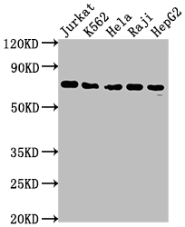
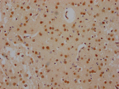
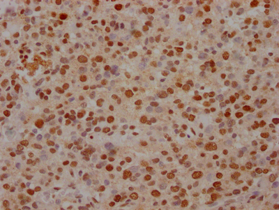
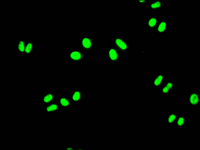
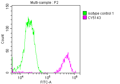
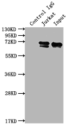
Validation testing demonstrates robust performance across multiple experimental platforms. Western blot analysis detects a clean band at the expected 69 kDa molecular weight across diverse human cell lines including Jurkat, K562, HeLa, Raji, and HepG2, confirming reliable detection in both suspension and adherent cell types. Immunohistochemistry staining has been validated in paraffin-embedded human brain tissue and glioma cancer samples using citrate buffer antigen retrieval, providing researchers studying neural biology or CNS malignancies with a validated detection tool. Immunofluorescence analysis in HeLa cells reveals clear nuclear localization consistent with FUBP1's known function as a transcription factor. Flow cytometry and immunoprecipitation applications have also been validated in Jurkat cells, offering flexibility for researchers investigating FUBP1 protein interactions or expression levels at the single-cell level.
This antibody supports investigations into epigenetics, nuclear signaling, and oncogene regulation where precise FUBP1 detection is essential.
There are currently no reviews for this product.

