IGHM Recombinant Monoclonal Antibody

Code CSB-RA582103A0HU
Size US$210
Order now
Image
  • IHC image of CSB-RA582103A0HU diluted at 1:100 and staining in paraffin-embedded human tonsil tissue performed on a Leica BondTM system. After dewaxing and hydration, antigen retrieval was mediated by high pressure in a citrate buffer (pH 6.0). Section was blocked with 10% normal goat serum 30min at RT. Then primary antibody (1% BSA) was incubated at 4°C overnight. The primary is detected by a Goat anti-rabbit polymer IgG labeled by HRP and visualized using 0.05% DAB.
  • IHC image of CSB-RA582103A0HU diluted at 1:100 and staining in paraffin-embedded human appendix tissue performed on a Leica BondTM system. After dewaxing and hydration, antigen retrieval was mediated by high pressure in a citrate buffer (pH 6.0). Section was blocked with 10% normal goat serum 30min at RT. Then primary antibody (1% BSA) was incubated at 4°C overnight. The primary is detected by a Goat anti-rabbit polymer IgG labeled by HRP and visualized using 0.05% DAB.
  • Immunofluorescence staining of RAJI cell with CSB-RA582103A0HU at 1:50 ,counter-stained with DAPI. The cells were fixed in 4% formaldehyde and blocked in 10% normal Goat Serum. The cells were then incubated with the antibody overnight at 4°C. The secondary antibody was Fluorescein Alexa Fluor 488-congugated AffiniPure Goat Anti-Rabbit IgG(H+L).
Have Questions? Leave a Message or Start an on-line Chat

Product Details

Uniprot No.
Target Names
IGHM
Alternative Names
Immunoglobulin heavy constant mu (Ig mu chain C region) (Ig mu chain C region BOT) (Ig mu chain C region GAL) (Ig mu chain C region OU), IGHM
Species Reactivity
Human
Immunogen
A synthesized peptide derived from Human IGHM protein
Immunogen Species
Homo sapiens (Human)
Conjugate
Non-conjugated
Clonality
Monoclonal
Isotype
Rabbit IgG
Clone No.
4D1
Purification Method
Affinity-chromatography
Concentration
It differs from different batches. Please contact us to confirm it.
Buffer
Rabbit IgG in 10mM phosphate buffered saline , pH 7.4, 150mM sodium chloride, 0.05% BSA, 0.02% sodium azide and 50% glycerol.
Form
Liquid
Tested Applications
ELISA, IHC, IF
Recommended Dilution
Application Recommended Dilution
IHC 1:50-1:200
IF 1:20-1:200
Troubleshooting and FAQs
Storage
Upon receipt, store at -20°C or -80°C. Avoid repeated freeze.
Lead Time
Basically, we can dispatch the products out in 1-3 working days after receiving your orders. Delivery time maybe differs from different purchasing way or location, please kindly consult your local distributors for specific delivery time.
Usage
For Research Use Only. Not for use in diagnostic or therapeutic procedures.

Customer Reviews and Q&A

 Customer Reviews

There are currently no reviews for this product.

Submit a Review here

Target Background

Function
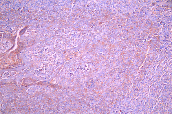
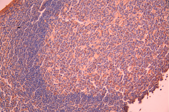
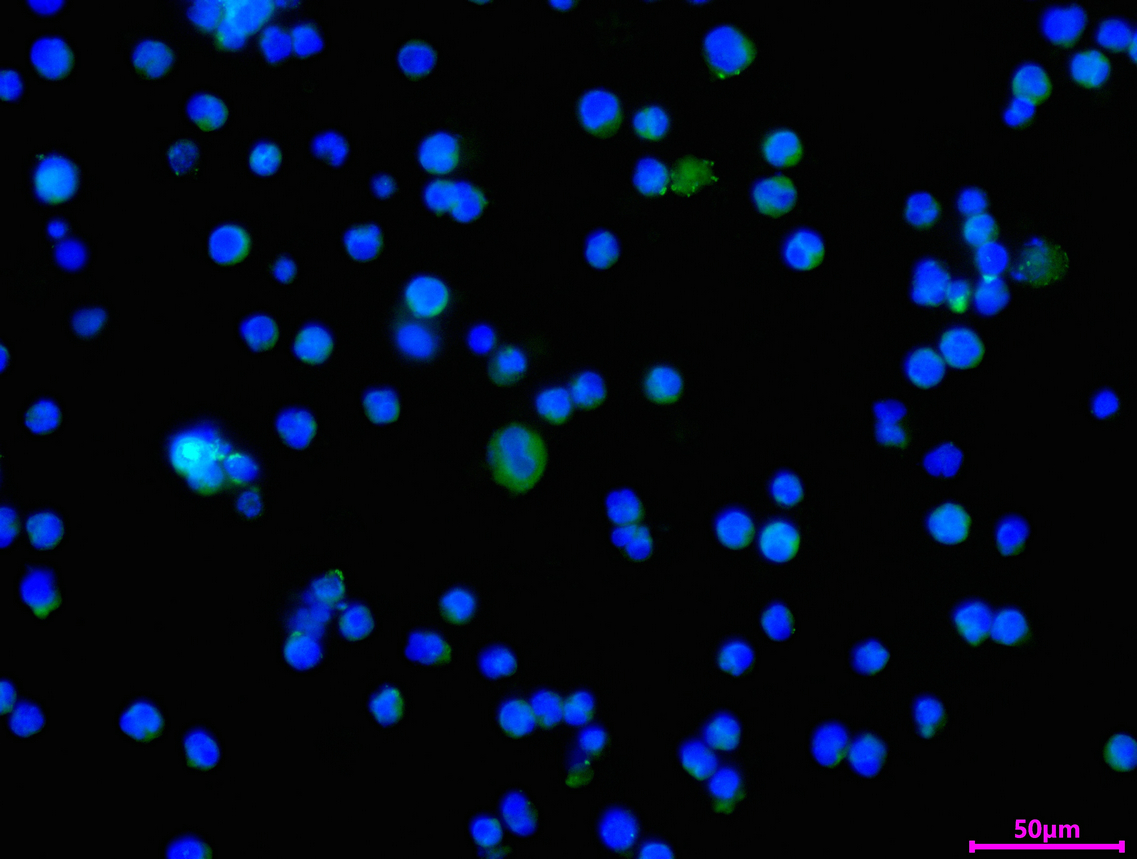
Constant region of immunoglobulin heavy chains. Immunoglobulins, also known as antibodies, are membrane-bound or secreted glycoproteins produced by B lymphocytes. In the recognition phase of humoral immunity, the membrane-bound immunoglobulins serve as receptors which, upon binding of a specific antigen, trigger the clonal expansion and differentiation of B lymphocytes into immunoglobulins-secreting plasma cells. Secreted immunoglobulins mediate the effector phase of humoral immunity, which results in the elimination of bound antigens. The antigen binding site is formed by the variable domain of one heavy chain, together with that of its associated light chain. Thus, each immunoglobulin has two antigen binding sites with remarkable affinity for a particular antigen. The variable domains are assembled by a process called V-(D)-J rearrangement and can then be subjected to somatic hypermutations which, after exposure to antigen and selection, allow affinity maturation for a particular antigen. IgM antibodies play an important role in primary defense mechanisms. They have been shown to be involved in early recognition of external invaders like bacteria and viruses, cellular waste and modified self, as well as in recognition and elimination of precancerous and cancerous lesions. The membrane-bound form is found in the majority of normal B-cells alongside with IgD. Membrane-bound IgM induces the phosphorylation of CD79A and CD79B by the Src family of protein tyrosine kinases. It may cause death of cells by apoptosis. It is also found in soluble form, which represents about 30% of the total serum immunoglobulins where it is found almost exclusively as a homopentamer. After the antigen binds to the B-cell receptor, the secreted form is secreted in large amounts.
Involvement in disease
Agammaglobulinemia 1, autosomal recessive (AGM1)
Subcellular Location
[Isoform 1]: Secreted. Note=During differentiation, B-lymphocytes switch from expression of membrane-bound IgM to secretion of IgM.; [Isoform 2]: Cell membrane; Single-pass type I membrane protein.
Database Links

HGNC: 5541

OMIM: 147020

UniGene: Hs.510635

icon of phone
Call us
301-363-4651 (Available 9 a.m. to 5 p.m. CST from Monday to Friday)
icon of address
Address
7505 Fannin St., Ste 610, Room 7 (CUBIO Innovation Center), Houston, TX 77054, USA
icon of social media
Join us with

Subscribe newsletter

Leave a message

* To protect against spam, please pass the CAPTCHA test below.
CAPTCHA verification
© 2007-2026 CUSABIO TECHNOLOGY LLC All rights reserved. 鄂ICP备15011166号-1
Place an order now

I. Product details

*
*
*
*

II. Contact details

*
*

III. Ship To

*
*
*
*
*
*
*

IV. Bill To

*
*
*
*
*
*
*
*