
Call us
301-363-4651 (Available 9 a.m. to 5 p.m. CST from Monday to Friday)

The NECTIN family comprises a group of cell adhesion molecules belonging to the immunoglobulin superfamily (IgSF), participating in essential processes such as cell-cell recognition, junction formation, and tissue architecture maintenance. Unlike classical cadherins, which primarily rely on calcium ions to mediate cell-cell junctions, the NECTIN family engages in more complex intercellular communication through diverse homophilic and heterophilic binding modes.
With advancing research, the functions of the NECTIN family have gradually expanded from traditional cell adhesion to nervous system development, viral infection, immune regulation, and tumor initiation and progression. Although different members share similar structural foundations, they play distinct roles in physiological and pathological processes due to differences in expression patterns, binding partners, and downstream signaling networks. Therefore, a systematic understanding of the structural characteristics and functional diversification of the NECTIN family is of great significance for deciphering cell-cell interaction mechanisms and developing relevant research tools and therapeutic strategies.
The NECTIN family typically includes two related subgroups: Nectin and Nectin-like (Necl), comprising a total of nine members. The Nectin subgroup includes Nectin-1, Nectin-2, Nectin-3, and Nectin-4; the Necl subgroup includes Necl-1, Necl-2, Necl-3, Necl-4, and Necl-5. Each member has multiple names used across different research fields. The table below summarizes the commonly used aliases (including protein names, former gene symbols, and other database accession numbers) of each member to facilitate literature retrieval.
| Family Member | Aliases |
|---|---|
| Nectin-1 | CD111, HVEC, HLGR, PRR1, PVRL1 |
| Nectin-2 | CD112, HVEB, PRR2, PVRL2, PVRR2 |
| Nectin-3 | CD113, PPR3, PRR3, PVRL3, PVRR3 |
| Nectin-4 | LNIR, PVRL4, PRR4 |
| Necl-1 | CADM3, SYNCAM3, IGSF4B, TSLL1 |
| Necl-2 | CADM1, SYNCAM1, IGSF4, IGSF4A, TSLC1 |
| Necl-3 | CADM2, SYNCAM2, IGSF4D |
| Necl-4 | CADM4, SYNCAM4, IGSF4C, TSLL2 |
| Necl-5 | CD155, HVED, PVR, PVS, TAGE4 |
From an evolutionary and structural perspective, Nectin and Necl belong to the same related molecular family; however, the two subgroups exhibit significant differences in intracellular structure and signal connection modes, which constitute an important basis for their further functional diversification.
All NECTIN family members are single-pass transmembrane proteins whose basic structure consists of an extracellular region, a transmembrane region, and an intracellular region.
Most members possess a typical immunoglobulin-like structure in the extracellular region, usually comprising one IgV domain and two IgC2 domains. This region is responsible for mediating intercellular binding and serves as the primary basis for homophilic and heterophilic interactions within the NECTIN family.
Unlike cadherins, which depend on calcium ions to maintain cell-cell junctions, binding between NECTIN molecules relies primarily on spatial complementarity among immunoglobulin-like domains. Consequently, the NECTIN family can form more flexible recognition patterns between different cell types [1,2].
The intracellular region determines how different members participate in cellular signal regulation. The cytoplasmic tails of classical Nectin members can bind the adaptor protein Afadin, which in turn connects to the actin cytoskeleton, converting adhesion events at the cell surface into cytoskeletal rearrangements and signal responses [2].
In contrast, Necl members generally do not function through the classical Afadin pathway; instead, they participate in cell junctions, migration, and signal regulation through other intracellular binding proteins. This structural difference enables NECTIN family members, despite sharing similar extracellular structures, to engage in distinct biological processes.
One of the most important features of the NECTIN family is the ability of its members to form multiple binding modes.
Some members can form stable junctions through homophilic binding, such as mutual recognition between identical molecules. At the same time, heterophilic binding can also occur between different members, for example, the interaction between Nectin-1 and Nectin-3, and the heterodimerization between Necl-4 and Necl-1 [17].
This heterophilic binding capacity distinguishes the NECTIN family from many traditional adhesion molecules. The combination of different NECTIN members expressed on the cell surface can determine whether cell-cell contact occurs and what type of junction is formed, thereby supporting complex cellular arrangement and spatial organization during tissue development.
In the nervous system, precise cell pairing is particularly critical for synapse formation. The heterophilic binding characteristics of NECTIN molecules can help different nerve cells establish selective contacts, providing a molecular foundation for synaptic structure formation [3].
One of the most important functions of classical Nectin members is their participation in junction formation through Afadin.
When Nectin molecules on the cell surface bind to each other, their intracellular regions can recruit Afadin. As an important scaffold protein linking membrane proteins and the actin network, Afadin can promote cytoskeletal reorganization and participate in the maturation of adherens junctions.
Studies have found that Nectin-mediated junctions do not exist independently but function in concert with other cell adhesion systems. In epithelial cells, the Nectin–Afadin complex can promote the formation of E-cadherin-mediated adherens junctions and participate in the establishment of tight junctions. Therefore, the NECTIN family plays a "coordinator" role in tissue architecture maintenance rather than simply providing mechanical connections.
With advancing research, the NECTIN family has been found to influence diverse cellular behaviors, including migration, proliferation, polarity establishment, and cell differentiation.
Cell adhesion itself is also a signaling event. When NECTIN molecules on the cell surface bind to adjacent cells, they can alter cytoskeletal status through intracellular scaffold proteins and signaling molecules, further influencing cell movement and morphological changes.
For example, in addition to participating in cell adhesion, Necl-5/CD155 is also involved in the regulation of cell migration and proliferation. In normal cells, CD155 participates in modulating cell motility; in tumor cells, its aberrant expression may enhance migratory capacity and further influence disease progression [4].
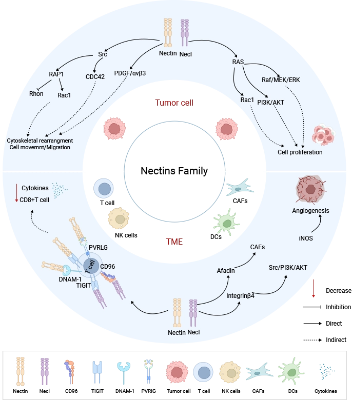
Figure 1. Nectin Family-Mediated Oncogenic Signaling in Tumor Cells and the Tumor Microenvironment [2]
Although NECTIN family members share similar structural foundations, they exhibit significant differences in tissue expression, binding partners, and biological functions. Among them, Nectin-1, Nectin-2/CD112, Nectin-4, and Necl-5/CD155 are the most intensively studied members, whereas CADM family members are primarily investigated in the context of the nervous system and tumors.
Nectin-1 (CD111) is one of the earliest discovered and most thoroughly studied Nectin members, widely expressed in epithelial tissues, the nervous system, and embryo development-related tissues. Its primary function is to participate in tissue architecture formation by mediating intercellular recognition and junctions.
During development, Nectin-1 is involved in cellular arrangement and morphogenesis across multiple tissues. Human genetic studies have found that mutations in the NECTIN1 gene can cause cleft lip/palate-ectodermal dysplasia syndrome (CLPED1), with patients exhibiting phenotypes such as cleft lip/palate, dental anomalies, and ectodermal tissue developmental defects [5]. This finding demonstrates that NECTIN1-mediated cell junctions are not only structurally significant but also indispensable regulatory factors in normal tissue development.
In addition to its role in tissue development, Nectin-1 holds important virological significance. Alphaherpesviruses such as herpes simplex virus type 1/2 (HSV-1/2) and pseudorabies virus (PRV) can exploit Nectin-1 as an important receptor for host cell entry, achieving viral-cell membrane fusion through binding of viral envelope glycoproteins to the Nectin-1 extracellular domain [6]. This discovery has made Nectin-1 an important molecule for studying viral infection mechanisms and host cell recognition.
Thus, Nectin-1 exemplifies a typical characteristic of the NECTIN family: the same cell surface adhesion molecule can participate in normal intercellular communication while also being exploited by pathogens to accomplish infection.
Nectin-2 (CD112) is currently one of the most immunologically valuable members of the NECTIN family.
Early research primarily focused on the role of CD112 in cell junctions. Subsequent studies revealed that CD112 also serves as a ligand for multiple immune receptors, including CD226 (DNAM-1), TIGIT, and PVRIG, making it an important molecule linking cell surface recognition and immune regulation [7,8].
CD226 is an activating receptor predominantly expressed on natural killer (NK) cells and a subset of T cells. Binding of CD112 to CD226 can promote the recognition and killing of target cells by immune cells. However, CD112 can also bind the inhibitory receptor PVRIG and participate in the inhibition of T cell and NK cell function.
Therefore, CD112 does not possess a simple "activating" or "inhibitory" attribute; rather, it sits within a dynamic regulatory network. Competition among different receptors for CD112 binding can influence the direction of signals ultimately received by immune cells.
In the tumor microenvironment, tumor cells may influence immune cell recognition by altering CD112 expression levels. In recent years, research surrounding the CD112–PVRIG axis has gradually increased, and this pathway is considered a potential novel immune regulatory direction following PD-1/PD-L1 and TIGIT.
Compared with Nectin-2 and Nectin-4, Nectin-3 has received less attention in disease research, yet its role in cell recognition within specific tissues is of significant importance.
The most prominent feature of Nectin-3 is its ability to form highly selective heterophilic binding with other Nectin members. For example, the interaction between Nectin-1 and Nectin-3 is believed to participate in cell recognition processes between nerve cells and within synaptic structures.
Cellular junctions in the nervous system are highly specific. Different types of nerve cells need to form stable connections at correct locations, and simple mechanical adhesion cannot meet such refined demands. Nectin-3 provides a molecular recognition mechanism for neural network formation through specific combinations with other members.
In addition, Nectin-3 participates in cell junctions within the reproductive system. Studies have shown that interactions among NECTIN family members are involved in maintaining junctions between Sertoli cells and germ cells during spermatogenesis [9].
At present, Nectin-3 has not yet become a clear drug development target, but it holds significant value for understanding tissue-specific cell recognition mechanisms.
Nectin-4 (PVRL4) is currently the most clinically successful member of the NECTIN family.
Initially discovered in relation to embryonic tissue development, subsequent studies found that Nectin-4 is aberrantly expressed in a variety of tumor tissues, including urothelial carcinoma, breast cancer, lung cancer, and pancreatic cancer [10].
Compared with normal adult tissues, Nectin-4 expression is markedly elevated in some tumor cells. Additionally, its cell surface localization provides favorable antibody accessibility. These characteristics have made Nectin-4 an important candidate target for antibody drug development.
The most representative application to date is the Nectin-4-targeted antibody-drug conjugate (ADC), Enfortumab vedotin. This drug utilizes an anti-Nectin-4 antibody to recognize Nectin-4 on the tumor cell surface, and upon internalization, releases the cytotoxic payload MMAE, thereby exerting antitumor effects [11].
Clinical studies have demonstrated clear therapeutic efficacy of Enfortumab vedotin in patients with advanced urothelial carcinoma, and it has received regulatory approval for relevant indications [12]. Recently, the FDA further approved its combination with pembrolizumab for perioperative therapy in muscle-invasive bladder cancer (MIBC), regardless of cisplatin eligibility.
The successful validation of Nectin-4 is of great significance: it proves that cell adhesion molecules can serve not only as objects of disease mechanism research but also as direct drug development targets. Current research surrounding Nectin-4 has shifted from "whether it can be targeted" to how to optimize therapeutic outcomes, including expression heterogeneity, resistance mechanisms, and combination therapeutic strategies.
The gene names for Necl-1 to Necl-4 are CADM3, CADM1, CADM2, and CADM4, respectively. It is worth noting that Necl-5 is not classified within the CADM family but is an independent member parallel to classical Nectin molecules. Compared with classical Nectin members, CADM family research is more concentrated in the nervous system and tumor-related fields.
Among them, CADM1 (Necl-2) is one of the most intensively studied members. CADM1 participates in intercellular adhesion and plays a role in synapse formation in the nervous system.
Meanwhile, multiple studies have found that abnormal CADM1 expression is associated with various tumors; particularly in lung cancer, decreased CADM1 expression correlates with reduced cell adhesion capacity and enhanced tumor invasion [13].
CADM2 is predominantly expressed in the nervous system and is believed to participate in information exchange between nerve cells and maintenance of neural functions [14]. CADM3 and CADM4 are also involved in cell junctions within neural tissues and have received attention in some tumor studies.
Overall, CADM members embody the characteristics of NECTIN-like molecules: they do not exist merely as structural adhesion molecules but influence tissue function by regulating intercellular communication. However, compared with Nectin-4 and CD155, the CADM family has not yet established a clear clinical translation direction.
Necl-5 (CD155) is one of the most functionally complex members of the NECTIN family.
CD155 was first discovered as the poliovirus receptor. Viruses can exploit CD155 to mediate cell entry, making it a classic subject of virus-host interaction research [15].
Subsequent studies found that CD155 is also involved in normal cell migration, proliferation, and cytoskeletal regulation. During cell migration, CD155 can influence cell movement-related signals, and in various tumors, elevated CD155 expression is associated with enhanced invasiveness [4].
In recent years, the hottest research area for CD155 has been tumor immunity. CD155 can bind multiple immune receptors, including TIGIT, CD226, and CD96. Among these, CD226 generally mediates immune activation, TIGIT has a clearly defined immunosuppressive effect, while CD96 also exerts immunosuppressive functions in most contexts, although its specific mechanisms are still under intensive investigation [16].
Because the same ligand simultaneously participates in both activating and inhibitory signal regulation, CD155 has become a critical balancing node in tumor immunity. When tumor cells highly express CD155, they may enhance inhibitory pathway signals such as TIGIT, restricting immune cell function.
Therefore, Necl-5 (CD155) and Nectin-4 represent two distinct value models of the NECTIN family in the tumor field: Nectin-4 primarily serves as a tumor cell surface antigen for direct drug delivery; whereas CD155 acts as an immune regulatory network node influencing immune cell function, representing an important potential target for tumor immunotherapy.
Research on the NECTIN family involves protein-protein interactions, cellular signaling, and disease mechanisms, requiring diverse experimental tools. CUSABIO provides a comprehensive portfolio of research products covering NECTIN family members, applicable to protein-protein interaction analysis, expression detection, receptor binding functional validation, and other scenarios, supporting basic scientific research and drug development.
| Target | Code | Product Name | Source |
|---|---|---|---|
| PVR | CSB-CF019093HU(A4) | Recombinant Human Poliovirus receptor (PVR) | in vitro E.coli expression system |
| PVR | CSB-EP019093HU | Recombinant Human Poliovirus receptor (PVR), partial | E.coli |
| NECTIN2 | CSB-EP835690HU | Recombinant Human Nectin-2 (NECTIN2), partial | E.coli |
| PVRL4 | CSB-YP822274HU | Recombinant Human Poliovirus receptor-related protein 4 (NECTIN4), partial | Yeast |
| NECTIN2 | CSB-AP005081HU | Recombinant Human Nectin-2 (NECTIN2), partial (Active) | Mammalian cell |
| NECTIN4 | CSB-AP005571HU | Recombinant Human Nectin-4 (NECTIN4), partial (Active) | Mammalian cell |
| NECTIN4 | CSB-MP822274HU | Recombinant Human Nectin-4 (NECTIN4), partial (Active) | Mammalian cell |
| PVR | CSB-MP019093HU(M) | Recombinant Human Poliovirus receptor (PVR) (I340M), partial (Active) | Mammalian cell |
| PVR | CSB-MP019093HUc7 | Recombinant Human Poliovirus receptor (PVR), partial | Mammalian cell |
| Cadm1 | CSB-MP004425MO | Recombinant Mouse Cell adhesion molecule 1 (Cadm1), partial (Active) | Mammalian cell |
| CADM1 | CSB-MP004425HU | Recombinant Human Cell adhesion molecule 1 (CADM1), partial (Active) | Mammalian cell |
| CADM1 | CSB-MP004425HUd9 | Recombinant Human Cell adhesion molecule 1 (CADM1), partial | Mammalian cell |
| NECTIN2 | CSB-MP5776MOV | Recombinant Macaca fascicularis Nectin cell adhesion molecule 2 (NECTIN2), partial (Active) | Mammalian cell |
| NECTIN1 | CSB-MP621867HU-B | Recombinant Human Nectin-1(NECTIN1), partial, Biotinylated | Mammalian cell |
| NECTIN2 | CSB-MP835690HUd7 | Recombinant Human Nectin-2 (NECTIN2), partial | Mammalian cell |
| NECTIN4 | CSB-EP822274HU-B | Recombinant Human Nectin-4 (NECTIN4), partial, Biotinylated | E.coli |
| NECTIN2 | CSB-MP835690HU | Recombinant Human Nectin-2 (NECTIN2), partial | Mammalian cell |
| Target | Code | Product Name | Tested Applications |
|---|---|---|---|
| PVR | CSB-RA567944A0HU | PVR Recombinant Monoclonal Antibody | ELISA, WB, IHC |
| NECTIN2 | CSB-RA835690MA1HU | NECTIN2 Recombinant Monoclonal Antibody | ELISA |
| PVRL2 | CSB-RA582013A0HU | PVRL2 Recombinant Monoclonal Antibody | ELISA, IHC |
| CADM2 | CSB-PA003005 | CADM2 Antibody | WB, IHC, IF, ELISA |
| NECTIN3 | CSB-PA003390 | PVRL3 Antibody | WB, ELISA |
| NECTIN3 | CSB-PA005242 | PVRL3 Antibody | WB, ELISA |
| CADM4 | CSB-PA008004 | CADM4 Antibody | WB, IHC, IF, ELISA |
| CADM3 | CSB-PA040111 | CADM3 Antibody | WB, IHC, IF, ELISA |
| NECTIN4 | CSB-PA060216 | PVRL4 Antibody | WB, ELISA |
| CADM1 | CSB-PA004340 | CADM1 Antibody | WB, IF, ELISA |
| NECTIN1 | CSB-PA006247 | PVRL1 Antibody | WB, ELISA |
| NECTIN2 | CSB-PA006259 | PVRL2 Antibody | WB, ELISA |
| PVR | CSB-PA006288 | PVR Antibody | WB, ELISA |
| CADM1 | CSB-PA004425GA01HU | CADM1 Antibody | ELISA, WB |
| CADM3 | CSB-PA004427GA01HU | CADM3 Antibody | ELISA, WB |
| NECTIN2 | CSB-PA019096GA01HU | PVRL2 Antibody | ELISA, WB |
| NECTIN3 | CSB-PA019097GA01HU | PVRL3 Antibody | ELISA, WB, IHC |
| CADM3 | CSB-PA130108 | CADM3 Antibody | ELISA, IHC |
| CADM3 | CSB-PA195475 | CADM3 Antibody | ELISA, IHC |
| PVR | CSB-PA297000 | PVR Antibody | ELISA, WB, IHC |
| Target | Code | Product Name | Detection Range | Sensitivity |
|---|---|---|---|---|
| PVR | CSB-EL019093HU | Human Poliovirus receptor(PVR) ELISA kit | serum, plasma, tissue homogenates | 7.8 ng/mL-500 ng/mL |
[1] de Agustín-Durán D,Mateos-White I,Fabra-Beser J, et al. Stick around: Cell-Cell Adhesion Molecules during Neocortical Development. Cells. 2021;10 (1).
[2] Chen R,Chen R,Chen W, et al. Nectins: Orchestrating tumor progression and immune evasion. Crit Rev Oncol Hematol. 2026;217:105058.
[3] Tomorsky J,Parker PRL,Doe CQ, et al. Precise levels of nectin-3 are required for proper synapse formation in postnatal visual cortex. Neural Dev. 2020;15 (1):13.
[4] Paolini R,Molfetta R. CD155 and Its Receptors as Targets for Cancer Therapy. Int J Mol Sci. 2023;24 (16).
[5] Lough KJ,Lough KJ,Spitzer DC, et al. Disruption of the nectin-afadin complex recapitulates features of the human cleft lip/palate syndrome CLPED1. Development. 2020;147 (21).
[6] Sayers CL,Elliott G. Herpes Simplex Virus 1 Enters Human Keratinocytes by a Nectin-1-Dependent, Rapid Plasma Membrane Fusion Pathway That Functions at Low Temperature. J Virol. 2016;90 (22):10379-10389.
[7] Sanchez-Correa B,Valhondo I,Hassouneh F, et al. DNAM-1 and the TIGIT/PVRIG/TACTILE Axis: Novel Immune Checkpoints for Natural Killer Cell-Based Cancer Immunotherapy. Cancers (Basel). 2019;11 (6).
[8] Buckle I,Guillerey C. Inhibitory Receptors and Immune Checkpoints Regulating Natural Killer Cell Responses to Cancer. Cancers (Basel). 2021;13 (17).
[9] Li T,Yao J,Zhang Q, et al. Chronic stress impairs male spermatogenesis function and Nectin-3 protein expression in the testis. Physiol Res. 2020;69 (2):297-306.
[10] Ganguli N,Mukherjee S,Samanta D. Membrane glycoprotein nectin-4 in tumor associated pathways and anti-cancer therapeutics. Int J Biol Macromol. 2025;330 (Pt 2):148023.
[11] Shostak Y,Nadell R,Capo L, et al. Enfortumab Vedotin Antibody-Drug Conjugate Targeting Nectin-4 Is a Highly Potent Therapeutic Agent in Multiple Preclinical Cancer Models. Cancer Res. 2016;76 (10):3003-13.
[12] Powles T,Rosenberg JE,Sonpavde GP, et al. Enfortumab Vedotin in Previously Treated Advanced Urothelial Carcinoma. N Engl J Med. 2021;384 (12):1125-1135.
[13] Li H,Gao J,Zhang S. Functional and Clinical Characteristics of Cell Adhesion Molecule CADM1 in Cancer. Front Cell Dev Biol. 2021;9:714298.
[14] Pasman JA,Chen Z,Smit DJA, et al. The CADM2 Gene and Behavior: A Phenome-Wide Scan in UK-Biobank. Behav Genet. 2022;52 (4-5):306-314.
[15] Gao J,Zheng Q,Xin N, et al. CD155, an onco-immunologic molecule in human tumors. Cancer Sci. 2017;108 (10):1934-1938.
[16] Kučan Brlić P,Lenac Roviš T,Cinamon G, et al. Targeting PVR (CD155) and its receptors in anti-tumor therapy. Cell Mol Immunol. 2019;16 (1):40-52.
[17] Liu X,Li D,Li C, et al. Structure of the heterophilic interaction between the nectin-like 4 and nectin-like 1 molecules. Proc Natl Acad Sci U S A. 2019;116 (6):2068-2077.


