Call us
301-363-4651 (Available 9 a.m. to 5 p.m. CST from Monday to Friday)
| Code | CSB-RA782379A0HU |
| Size | US$210 |
| Order now | |
| Image |
|
| Have Questions? | Leave a Message or Start an on-line Chat |
| Application | Recommended Dilution |
|---|---|
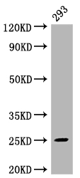
| WB | 1:500-1:5000 |
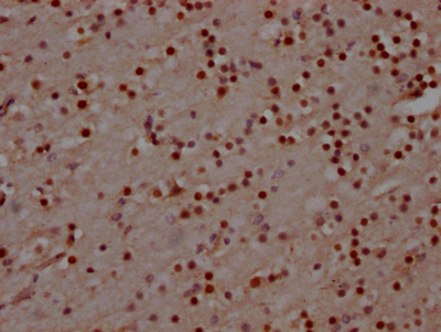
| IHC | 1:50-1:200 |
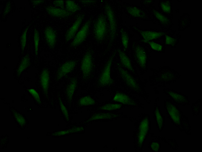
| IF | 1:20-1:200 |
MAD2L2, also known as MAD2B or REV7, serves as a critical regulator at the intersection of genome stability and cell cycle control. This protein functions as a component of the shieldin complex, protecting DNA double-strand breaks from excessive resection, while also participating in translesion DNA synthesis as part of the REV1-polymerase zeta complex. Its dual roles in DNA damage tolerance and repair make MAD2L2 a compelling target for researchers investigating cancer biology, particularly in contexts involving BRCA-deficient tumors and therapeutic resistance mechanisms.
This recombinant monoclonal antibody, clone 4D8, offers the reproducibility and consistency that demanding experimental workflows require. Because the antibody sequence is defined and produced recombinantly in rabbit host, researchers can expect reliable performance across experiments and between lots, eliminating the variability often encountered with traditional hybridoma-derived antibodies.
Validation data demonstrates robust performance across multiple applications. Western blot analysis of 293 whole cell lysate reveals a clean band at the predicted molecular weight of 25 kDa, confirming specific target recognition without apparent degradation products or non-specific binding. The antibody has also been validated for immunohistochemistry in paraffin-embedded human brain tissue, enabling researchers to examine MAD2L2 expression patterns in archival clinical specimens. For subcellular localization studies, immunofluorescence staining in HeLa cells provides clear visualization of the protein's distribution.
With validated applications spanning ELISA, western blot, immunohistochemistry, and immunofluorescence, this antibody supports diverse experimental approaches for investigating MAD2L2 function in DNA repair pathways, cell cycle checkpoint regulation, and cancer-related research programs.
There are currently no reviews for this product.

