MAS1L Recombinant Monoclonal Antibody

Code CSB-RA971033A0HU
Size US$210
Order now
Image
  • Western Blot
    Positive WB detected in: MCF-7 whole cell lysate, Hela whole cell lysate, A549 whole cell lysate, HepG2 whole cell lysate, PC-3 whole cell lysate, Jurkat whole cell lysate, Rat liver tissue
    All lanes: MAS1L antibody at 1:1000
    Secondary
    Goat polyclonal to rabbit IgG at 1/50000 dilution
    Predicted band size: 43 kDa
    Observed band size: 36-50 kDa
  • Immunofluorescence staining of MCF-7 cell with CSB-RA971033A0HU at 1:50, counter-stained with DAPI. The cells were fixed in 4% formaldehyde and blocked in 10% normal Goat Serum. The cells were then incubated with the antibody overnight at 4°C. The secondary antibody was Alexa Fluor 595-congugated AffiniPure Goat Anti-Rabbit IgG(H+L).
  • Immunofluorescence staining of Hela cell with CSB-RA971033A0HU at 1:50, counter-stained with DAPI. The cells were fixed in 4% formaldehyde and blocked in 10% normal Goat Serum. The cells were then incubated with the antibody overnight at 4°C. The secondary antibody was Alexa Fluor 595-congugated AffiniPure Goat Anti-Rabbit IgG(H+L).
  • Overlay Peak curve showing Hela cells stained with CSB-RA971033A0HU (red line) at 1:100. The cells were fixed in 4% formaldehyde and permeated by 0.2% TritonX-100. Then 10% normal goat serum to block non-specific protein-protein interactions followed by the antibody (1ug/1*106cells) for 45min at 4℃. The secondary antibody used was FITC-conjugated Goat Anti-Rabbit IgG(H+L) at 1:200 dilution for 35min at 4℃.Control antibody (green line) was Rabbit IgG (1ug/1*106cells) used under the same conditions.
Have Questions? Leave a Message or Start an on-line Chat
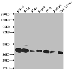
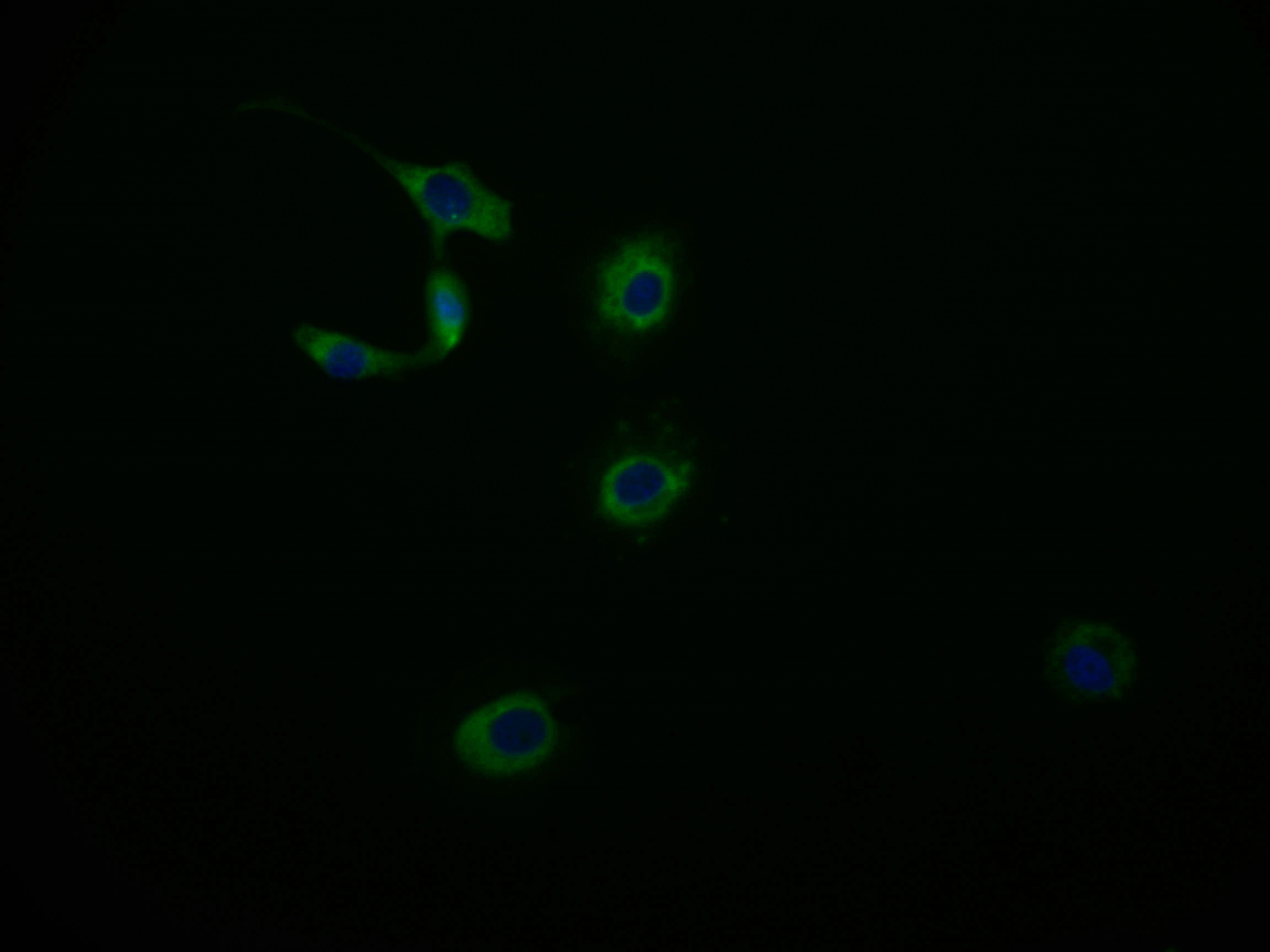
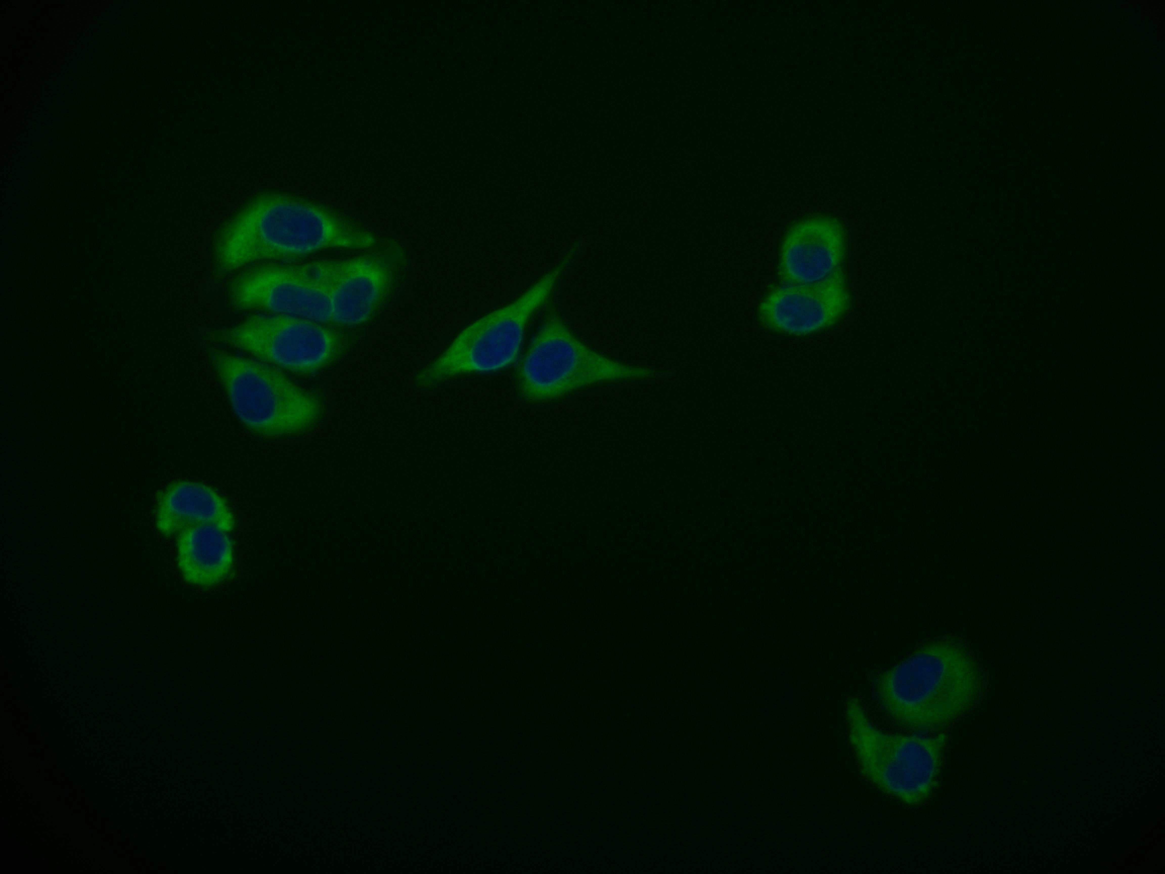
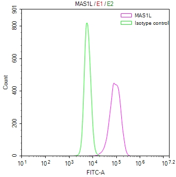
Product Details

Uniprot No.
Target Names
MAS1L
Alternative Names
Mas-related G-protein coupled receptor MRG (MAS-R) (MAS1-like), MAS1L, MRG
Species Reactivity
Human, Rat
Immunogen
A synthesized peptide derived from human MAS1L
Immunogen Species
Homo sapiens (Human)
Conjugate
Non-conjugated
Clonality
Monoclonal
Isotype
Rabbit IgG
Clone No.
15B8
Purification Method
Affinity-chromatography
Concentration
It differs from different batches. Please contact us to confirm it.
Buffer
Rabbit IgG in phosphate buffered saline, pH 7.4, 150mM NaCl, 0.02% sodium azide and 50% glycerol.
Form
Liquid
Tested Applications
ELISA, WB, IF, FC
Recommended Dilution
Application Recommended Dilution
WB 1:500-1:2000
IF 1:50-1:200
FC 1:50-1:200
Troubleshooting and FAQs
Storage
Upon receipt, store at -20°C or -80°C. Avoid repeated freeze.
Lead Time
Basically, we can dispatch the products out in 1-3 working days after receiving your orders. Delivery time maybe differs from different purchasing way or location, please kindly consult your local distributors for specific delivery time.
Description

The MAS1L recombinant monoclonal antibody is produced through a process of genetic engineering that involves cloning and expression of the gene encoding for the MAS1L monoclonal antibody. The immunogen used to generate the MAS1L monoclonal antibody is the synthesized peptide derived from human MAS1L protein. The obtained MAS1L recombinant monoclonal antibody is purified using affinity chromatography to ensure high and purity. This MAS1L recombinant monoclonal antibody can specifically recognize and bind to the MAS1L protein. It has been validated for use in human and rat samples. Multiple applications including ELISA, WB, IF, and FC have been carried out to test the quality and specificity of the MAS1L recombinant monoclonal antibody.

MAS1L is primarily expressed in the adrenal gland, but is also found in other tissues including the brain, kidney, and heart. It is involved in the regulation of blood pressure and cardiovascular homeostasis through the renin-angiotensin system (RAS). MAS1L is activated by angiotensin-(1-7), a peptide hormone that opposes the effects of angiotensin II, which promotes vasoconstriction and raises blood pressure. Activation of MAS1L leads to vasodilation and a decrease in blood pressure. MAS1L has been found to be expressed on immune cells such as macrophages, and its activation has been shown to inhibit the release of inflammatory cytokines.

Customer Reviews and Q&A

 Customer Reviews

There are currently no reviews for this product.

Submit a Review here

Target Background

Gene References into Functions
  1. Mas, MrgD, and MRG mediated Ang IV-stimulated AA release that was highest for Mas. While Ang III activated Mas and MrgX2, Ang II stimulated AA release via Mas and MRG. PMID: 18636314
Subcellular Location
Cell membrane; Multi-pass membrane protein.
Protein Families
G-protein coupled receptor 1 family, Mas subfamily
Database Links

HGNC: 13961

OMIM: 607235

KEGG: hsa:116511

STRING: 9606.ENSP00000366331

UniGene: Hs.533297

icon of phone
Call us
301-363-4651 (Available 9 a.m. to 5 p.m. CST from Monday to Friday)
icon of address
Address
7505 Fannin St., Ste 610, Room 7 (CUBIO Innovation Center), Houston, TX 77054, USA
icon of social media
Join us with

Subscribe newsletter

Leave a message

* To protect against spam, please pass the CAPTCHA test below.
CAPTCHA verification
© 2007-2024 CUSABIO TECHNOLOGY LLC All rights reserved. 鄂ICP备15011166号-1
Place an order now

I. Product details

*
*
*
*

II. Contact details

*
*

III. Ship To

*
*
*
*
*
*
*

IV. Bill To

*
*
*
*
*
*
*
*