Call us
301-363-4651 (Available 9 a.m. to 5 p.m. CST from Monday to Friday)
| Code | CSB-RA33245A2GMY |
| Size | US$420 |
| Order now | |
| Image |
|
| Have Questions? | Leave a Message or Start an on-line Chat |
| Application | Recommended Dilution |
|---|---|
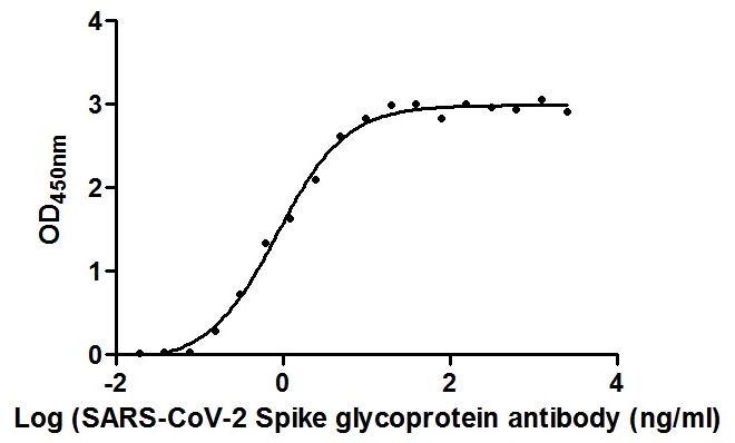
| ELISA | 1:10000-1:100000 |
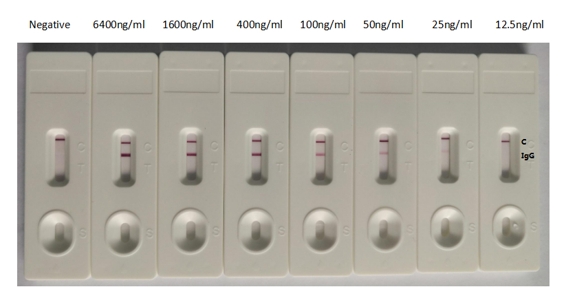
| GICA | 1:10000-1:40000 |
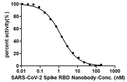
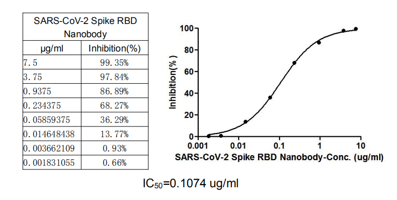
| Neutralising | 1:100-1:10000 |
The SARS-CoV-2 spike glycoprotein receptor-binding domain represents a critical interface in viral pathogenesis, mediating the initial attachment to human ACE2 receptors that enables cellular entry. This interaction has made the RBD a focal point for therapeutic development, diagnostic assay design, and fundamental studies of coronavirus biology.
This recombinant nanobody, derived from alpaca VHH sequences fused to a human IgG1 Fc domain, offers researchers a uniquely compact binding format with exceptional affinity. The recombinant production method ensures sequence-defined consistency between lots, eliminating the batch variability that can complicate longitudinal studies or assay standardization. Affinity chromatography purification delivers a reagent ready for demanding applications.
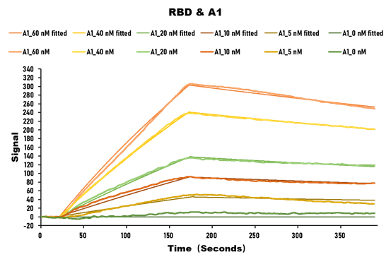
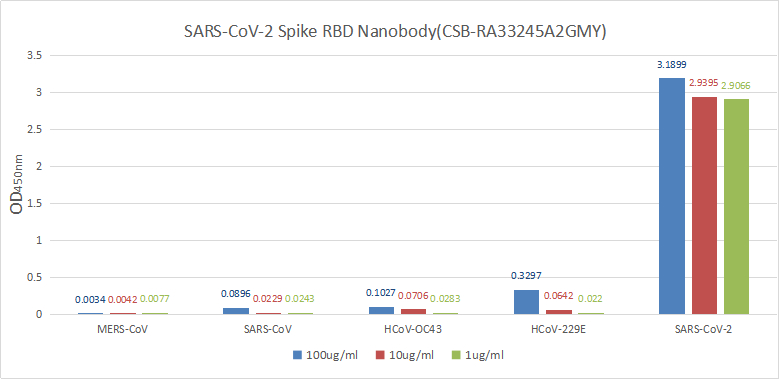
Validation data demonstrate impressive binding characteristics, with an EC50 of 0.8674 ng/ml in functional ELISA against immobilized S1-RBD and a measured affinity constant of 28.2 nM by surface plasmon resonance. Critically for neutralization studies, this nanobody effectively blocks the RBD-ACE2 interaction with an IC50 of 1.296 nM, providing a valuable tool for investigating viral entry mechanisms or developing competitive binding assays. Specificity testing confirms reactivity exclusively with SARS-CoV-2 S1-RBD, with no detectable binding to spike proteins from MERS-CoV, SARS-CoV, HCoV-OC43, or HCoV-229E, ensuring confidence in coronavirus-specific applications.
The antibody performs across multiple platforms including ELISA at dilutions from 1:10,000 to 1:100,000, colloidal gold immunochromatography assays with detection limits as low as 25 ng/ml, and neutralization studies. This versatility supports workflows ranging from high-throughput screening to rapid diagnostic development, making it a practical choice for virology research, vaccine evaluation studies, and infectious disease diagnostics.
There are currently no reviews for this product.
Where was CSB-RA33245A2GMY purified from? cell culture / hybridoma or serum / ascites?
What is the concentration of this antibody, do you have this antibody in stock?
I am interested in this product. What is the detection limit? Is it below 100-1000 SARS-CoV-2 particles?
Is there any advantage of Nanobody compared with traditional hybridoma monoclonal antibody?
What's the meaning of "Nanobody"?
What is the research significance of SARS-CoV-2 Spike RBD Nanobody?
In the Colloidal Gold Immunochromatography Assay, the detection limit was as low as 25ng/ml (1.75ng). Does it mean a concentration of 25ng/ml?

