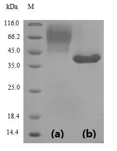
Recombinant Severe acute respiratory syndrome coronavirus 2 Spike glycoprotein (S), partial (Active)

In Stock
Code CSB-YP3324GMY1
Size $470
Order now
Image
  • (Tris-Glycine gel) Discontinuous SDS-PAGE (reduced) with 5% enrichment gel and 15% separation gel.
  • Activity
    Measured by its binding ability in a functional ELISA. Immobilized SARS-CoV-2-S1-RBD at 2 μg/ml can bind SARS-CoV-2-S Antibody (CSB-RA33245A1GMY), the EC50 of SARS-CoV-2-S1-RBD protein is 19.60-39.42 ng/ml. Biological Activity Assay
  • Activity
    Measured by its binding ability in a functional ELISA. Immobilized SARS-CoV-2-S1-RBD at 5 μg/ml can bind human ACE2 (CSB-MP866317HU), the EC50 of SARS-CoV-2-S1-RBD protein is 31.80 - 44.69 ng/ml. Biological Activity Assay
  • Activity
    Measured by its binding ability in a functional ELISA. Immobilized SARS-CoV-2-S1-RBD at 2 μg/ml can bind SARS-CoV-2-S Antibody (CSB-RA33245A0GMY), the EC50 of SARS-CoV-2-S1-RBD protein is 13.48-19.50 ng/ml. Biological Activity Assay
  • Activity
    SARS-CoV-2 Spike protein RBD his/sumostar tag (CSB-YP3324GMY1) captured on COOH chip can bind Human ACE2 protein Fc tag (CSB-MP866317HU) with an affinity constant of 100 nM as detected by LSPR Assay. Biological Activity Assay
  • Activity
    SARS-CoV-2 Spike protein RBD His/Sumostar Tag (CSB-YP3324GMY1) captured on COOH chip can bind SARS-CoV-2 Spike RBD Nanobody (CSB-RA33245A2GMY) with an affinity constant of 28.2nM as detected by LSPR Assay. Biological Activity Assay
Have Questions? Leave a Message or Start an on-line Chat

Product Details

Purity
Greater than 90% as determined by SDS-PAGE.
Endotoxin
Less than 1.0 EU/ug as determined by LAL method.
Activity
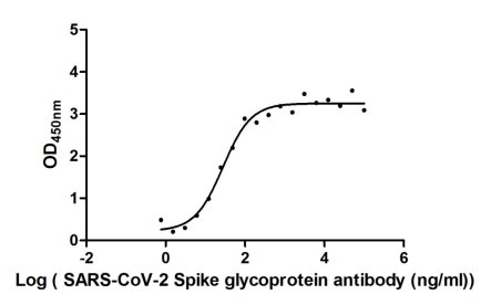
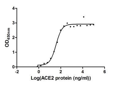
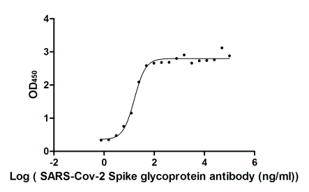
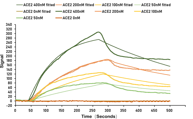
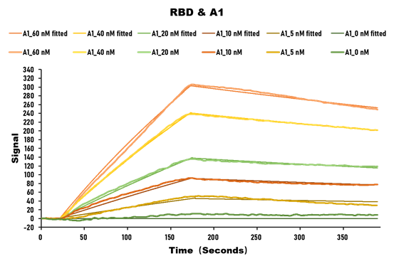
①Measured by its binding ability in a functional ELISA. Immobilized SARS-CoV-2-S1-RBD at 2 μg/ml can bind SARS-CoV-2-S Antibody (CSB-RA33245A1GMY), the EC50 of SARS-CoV-2-S1-RBD protein is 19.60-39.42 ng/ml. ②Measured by its binding ability in a functional ELISA. Immobilized SARS-CoV-2-S1-RBD at 5 μg/ml can bind human ACE2 (CSB-MP866317HU), the EC50 of SARS-CoV-2-S1-RBD protein is 31.80 - 44.69 ng/ml. ③Measured by its binding ability in a functional ELISA. Immobilized SARS-CoV-2-S1-RBD at 2 μg/ml can bind SARS-CoV-2-S Antibody (CSB-RA33245A0GMY), the EC50 of SARS-CoV-2-S1-RBD protein is 13.48-19.50 ng/ml. ④SARS-CoV-2 Spike protein RBD his/sumostar tag (CSB-YP3324GMY1) captured on COOH chip can bind Human ACE2 protein Fc tag (CSB-MP866317HU) with an affinity constant of 100 nM as detected by LSPR Assay. ⑤SARS-CoV-2 Spike protein RBD His/Sumostar Tag (CSB-YP3324GMY1) captured on COOH chip can bind SARS-CoV-2 Spike RBD Nanobody (CSB-RA33245A2GMY) with an affinity constant of 28.2nM as detected by LSPR Assay.
Target Names
S
Uniprot No.
Research Area
Microbiology
Alternative Names
S; 2; Spike glycoprotein; S glycoprotein; E2; Peplomer protein)
Species
Severe acute respiratory syndrome coronavirus 2 (2019-nCoV) (SARS-CoV-2)
Source
Yeast
Expression Region
319-541aa
Target Protein Sequence
RVQPTESIVRFPNITNLCPFGEVFNATRFASVYAWNRKRISNCVADYSVLYNSASFSTFKCYGVSPTKLNDLCFTNVYADSFVIRGDEVRQIAPGQTGKIADYNYKLPDDFTGCVIAWNSNNLDSKVGGNYNYLYRLFRKSNLKPFERDISTEIYQAGSTPCNGVEGFNCYFPLQSYGFQPTNGVGYQPYRVVVLSFELLHAPATVCGPKKSTNLVKNKCVNF
Mol. Weight
38.2 kDa
Protein Length
Partial
Tag Info
N-terminal 6xHis-sumostar-tagged
Form
Lyophilized powder
Note: We will preferentially ship the format that we have in stock, however, if you have any special requirement for the format, please remark your requirement when placing the order, we will prepare according to your demand.
Buffer
Lyophilized from a 0.2 μm sterile filtered 20 mM Tris-HCl, 0.5 M NaCl, 6% Trehalose, pH 8.0
Reconstitution
We recommend that this vial be briefly centrifuged prior to opening to bring the contents to the bottom. Please reconstitute protein in deionized sterile water to a concentration of 0.1-1.0 mg/mL.We recommend to add 5-50% of glycerol (final concentration) and aliquot for long-term storage at -20°C/-80°C. Our default final concentration of glycerol is 50%. Customers could use it as reference.
Troubleshooting and FAQs
Storage Condition
Store at -20°C/-80°C upon receipt, aliquoting is necessary for mutiple use. Avoid repeated freeze-thaw cycles.
Shelf Life
The shelf life is related to many factors, storage state, buffer ingredients, storage temperature and the stability of the protein itself.
Generally, the shelf life of liquid form is 6 months at -20°C/-80°C. The shelf life of lyophilized form is 12 months at -20°C/-80°C.
Lead Time
3-7 business days
Notes
Repeated freezing and thawing is not recommended. Store working aliquots at 4°C for up to one week.
Datasheet & COA
Please contact us to get it.

Customer Reviews and Q&A

 Customer Reviews
Average Rating:
5.0 - 2 reviews

Submit a Review here

Applications : Quantitative analysis

Review: Spearman’s rank correlation between ID50 and anti-RBD ELISA absolute quantification (μg/ml) in the total samples analyzed.

By Anonymous

Applications : ELISA assays

Review: In vitro immune response to partial SARS-CoV-2 spike glycoprotein in healthy controls and rheumatic disease patients stratified by anti-RBD IgG response (positive or negative).

By Anonymous

Target Background

Function
attaches the virion to the cell membrane by interacting with host receptor, initiating the infection. Binding to human ACE2 receptor and internalization of the virus into the endosomes of the host cell induces conformational changes in the Spike glycoprotein. Binding to host NRP1 and NRP2 via C-terminal polybasic sequence enhances virion entry into host cell. This interaction may explain virus tropism of human olfactory epithelium cells, which express high level of NRP1 and NRP2 but low level of ACE2. The stalk domain of S contains three hinges, giving the head unexpected orientational freedom. Uses human TMPRSS2 for priming in human lung cells which is an essential step for viral entry. Can be alternatively processed by host furin. Proteolysis by cathepsin CTSL may unmask the fusion peptide of S2 and activate membranes fusion within endosomes.; mediates fusion of the virion and cellular membranes by acting as a class I viral fusion protein. Under the current model, the protein has at least three conformational states: pre-fusion native state, pre-hairpin intermediate state, and post-fusion hairpin state. During viral and target cell membrane fusion, the coiled coil regions (heptad repeats) assume a trimer-of-hairpins structure, positioning the fusion peptide in close proximity to the C-terminal region of the ectodomain. The formation of this structure appears to drive apposition and subsequent fusion of viral and target cell membranes.; Acts as a viral fusion peptide which is unmasked following S2 cleavage occurring upon virus endocytosis.; May down-regulate host tetherin (BST2) by lysosomal degradation, thereby counteracting its antiviral activity.
Gene References into Functions
  1. Study presents crystal structure of C-terminal domain of SARS-CoV-2 (SARS-CoV-2-CTD) spike S protein in complex with human ACE2 (hACE2); hACE2-binding mode similar overall to that observed for SARS-CoV. However, details at the binding interface show that key residue substitutions in SARS-CoV-2-CTD slightly strengthen the interaction and lead to higher affinity for receptor binding than SARS-CoV receptor-binding domain. PMID: 32378705
  2. crystal structure of the receptor-binding domain (RBD) of the spike protein of SARS-CoV-2 bound to the cell receptor ACE2 PMID: 32365751
  3. crystal structure of the receptor-binding domain (RBD) of the spike protein of SARS-CoV-2 (engineered to facilitate crystallization) in complex with ACE2 PMID: 32320687
  4. Out of the two isolates from India compared to the isolates from Wuhan, China, one was found to harbor a mutation in its receptor-binding domain (RBD) at position 407 where, arginine was replaced by isoleucine. This mutation has been seen to change the secondary structure of the protein at that region and this can potentially alter receptor binding of the virus. PMID: 32275855
  5. Structural modeling of the SARS-CoV-2 spike glycoprotein show similar receptor utilization between SARS-CoV-2 and SARS-CoV, despite a relatively low amino acid similarity in the receptor binding module. Compared to SARS-CoV and all other coronaviruses in Betacoronavirus lineage B, an extended structural loop containing basic amino acids were identified at the interface of the receptor binding (S1) and fusion (S2) domains. PMID: 32245784
  6. crystal structure of CR3022, a neutralizing antibody from a SARS patient, in complex with the receptor-binding domain of the SARS-CoV-2 spike (S) protein to 3.1 A; study provides insight into how SARS-CoV-2 can be targeted by the humoral immune response and revealed a conserved, but cryptic epitope shared between SARS-CoV-2 and SARS-CoV PMID: 32225176
  7. SARS-CoV and SARS-CoV-2 spike proteins have comparable binding affinities achieved by balancing energetics and dynamics. The SARS-CoV-2-ACE2 complex contains a higher number of contacts, a larger interface area, and decreased interface residue fluctuations relative to the SARS-CoV-ACE2 complex. PMID: 32225175
  8. Interaction interface between cat/dog/pangolin/Chinese hamster ACE2 and SARS-CoV/SARS-CoV-2 S protein was simulated through homology modeling. Authors identified that N82 of ACE2 showed closer contact with receptor-binding domain of S protein than human ACE2. PMID: 32221306
  9. SARS-CoV-2 S glycoprotein harbors a furin cleavage site at the boundary between the S1/S2 subunits, which is processed during biogenesis and sets this virus apart from SARS-CoV and SARS-related CoVs; determined cryo-EM structures of the SARS-CoV-2 S ectodomain trimer. PMID: 32201080
  10. Study demonstrates that SARS-CoV-2 uses the SARS-CoV receptor ACE2 for entry and the serine protease TMPRSS2 for S protein priming. PMID: 32155444
  11. The ACE2-B0AT1 complex exists as a dimer of heterodimers. Structural alignment of the RBD-ACE2-B0AT1 ternary complex with the S protein of SARS-CoV-2 suggests that two S protein trimers can simultaneously bind to an ACE2 homodimer. PMID: 32142651
  12. study demonstrated SARS-CoV-2 S protein entry on 293/hACE2 cells is mainly mediated through endocytosis, and PIKfyve, TPC2 and cathepsin L are critical for virus entry; found that SARS-CoV-2 S protein could trigger syncytia in 293/hACE2 cells independent of exogenous protease; there was limited cross-neutralization activity between convalescent sera from SARS and COVID-19 patients PMID: 32132184
  13. study determined a 3.5-angstrom-resolution cryo-electron microscopy structure of the 2019-nCoV S trimer in the prefusion conformation; provided biophysical and structural evidence that the 2019-nCoV S protein binds angiotensin-converting enzyme 2 (ACE2) with higher affinity than does severe acute respiratory syndrome (SARS)-CoV S PMID: 32075877

Show More

Hide All

Subcellular Location
Virion membrane; Single-pass type I membrane protein. Host endoplasmic reticulum-Golgi intermediate compartment membrane; Single-pass type I membrane protein. Host cell membrane; Single-pass type I membrane protein.
Protein Families
Betacoronaviruses spike protein family
CUSABIO guaranteed quality
icon of phone
Call us
301-363-4651 (Available 9 a.m. to 5 p.m. CST from Monday to Friday)
icon of address
Address
7505 Fannin St., Ste 610, Room 7 (CUBIO Innovation Center), Houston, TX 77054, USA
icon of social media
Join us with

Subscribe newsletter

Leave a message

* To protect against spam, please pass the CAPTCHA test below.
CAPTCHA verification
© 2007-2024 CUSABIO TECHNOLOGY LLC All rights reserved. 鄂ICP备15011166号-1
webinars: DT3C facilitates antibody internalization X
Place an order now

I. Product details

*
*
*
*

II. Contact details

*
*

III. Ship To

*
*
*
*
*
*
*

IV. Bill To

*
*
*
*
*
*
*
*