Call us
301-363-4651 (Available 9 a.m. to 5 p.m. CST from Monday to Friday)
| Code | CSB-RA987582A0HU |
| Size | US$210 |
| Order now | |
| Image |
|
| Have Questions? | Leave a Message or Start an on-line Chat |
| Application | Recommended Dilution |
|---|---|
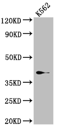
| WB | 1:500-1:5000 |
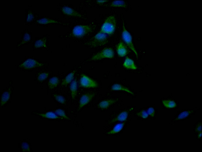
| IF | 1:20-1:200 |
SAE1, or SUMO-activating enzyme subunit 1, plays an essential role in the SUMOylation pathway, a post-translational modification system that regulates diverse cellular processes including transcription, DNA repair, cell cycle progression, and protein stability. As the regulatory subunit of the heterodimeric E1 activating enzyme, SAE1 works in concert with UBA2 to initiate the SUMO conjugation cascade, making it a critical node for researchers investigating protein modification networks and their implications in disease states.
This recombinant monoclonal antibody, clone 9D8, offers the reproducibility and consistency that demanding experimental workflows require. Because it is produced from a defined sequence in rabbit host cells, you can expect uniform performance across lots, eliminating the variability that can complicate long-term studies or multi-site collaborations. The antibody is raised against a synthetic peptide derived from human SAE1 and has been validated for human sample detection.
Western blot analysis demonstrates clean detection of SAE1 at the expected 39 kDa molecular weight in K562 whole cell lysate, with effective working dilutions ranging from 1:500 to 1:5000, giving you flexibility to optimize signal-to-background ratios for your specific experimental conditions. For cellular localization studies, immunofluorescence validation in HeLa cells confirms reliable staining performance at dilutions between 1:20 and 1:200, enabling visualization of SAE1 distribution patterns within fixed and permeabilized samples.
Whether you are exploring SUMOylation dynamics in cancer biology, investigating stress response pathways, or characterizing protein-protein interactions within the SUMO machinery, this antibody provides a dependable tool for advancing your cell biology research.
There are currently no reviews for this product.

