Call us
301-363-4651 (Available 9 a.m. to 5 p.m. CST from Monday to Friday)
| Code | CSB-MA000051M0m |
| Size | US$120 |
| Order now | |
| Image |
|
| Have Questions? | Leave a Message or Start an on-line Chat |
| Application | Recommended Dilution |
|---|---|
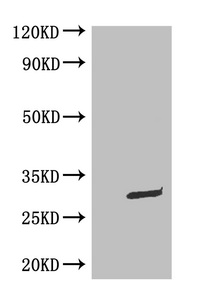
| WB | 1:500-1:5000 |
| IP | 1:200-1:2000 |
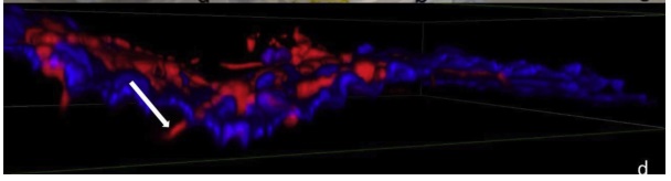
Applications : IHC
Sample dilution: 1: 500
Review: fluorescent microscopic analysis 12 h after inoculation showing the fungal hyphae expressing GFP (in red) penetrating the floral tissue (in blue).
By Anonymous
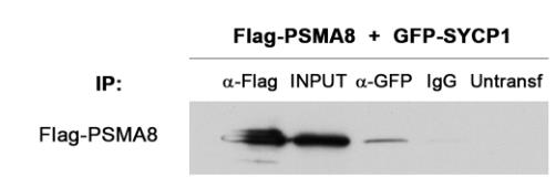
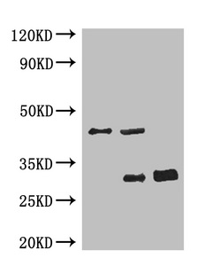
Applications : IP
Sample dilution: 1: 1000
Review: HEK293T cells were transfected with Flag-PSMA8 and GFP-SYCP1. Protein complexes were immunoprecipitated overnight with either an anti-Flag or anti-EGFP or IgGs (negative control), and were analyzed by immunoblotting with the indicated antibody. PSMA8 co-immunoprecipitates with SYCP1.
By Anonymous
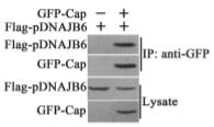
Applications : IP
Review: PCV2 Cap interacts with pDNAJB6 in transfected cells. HEK293T cells were co-transfected with GFP-Cap and Flag-pDNAJB6 expression plasmids or co-transfected with the pEGFP-N1 control vector and the Flag-pDNAJB6 expression plasmid. The cells transfected with pEGFP-N1 control vector, GFP-Cap expression plasmid or Flag-pDNAJB6 expression plasmid alone served as controls. The interaction of GFP-Cap with Flag-pDNAJB6 was identified through immunoprecipitation using anti-Flag antibodies or anti-GFP antibodies.
By Anonymous

