Recombinant Human Clarin-1 (CLRN1)

In Stock
Code CSB-CF005583HU
Abbreviation Recombinant Human CLRN1 protein
MSDS
Size $1620
Order now
Image
  • (Tris-Glycine gel) Discontinuous SDS-PAGE (reduced) with 5% enrichment gel and 15% separation gel.
Have Questions? Leave a Message or Start an on-line Chat

Product Details

Purity
Greater than 85% as determined by SDS-PAGE.
Activity
Not Test
Target Names
CLRN1
Uniprot No.
Research Area
Cancer
Species
Homo sapiens (Human)
Source
in vitro E.coli expression system
Expression Region
1-232aa
Target Protein Sequence
MPSQQKKIIFCMAGVFSFACALGVVTALGTPLWIKATVLCKTGALLVNASGQELDKFMGEMQYGLFHGEGVRQCGLGARPFRFSFFPDLLKAIPVSIHVNVILFSAILIVLTMVGTAFFMYNAFGKPFETLHGPLGLYLLSFISGSCGCLVMILFASEVKIHHLSEKIANYKEGTYVYKTQSEKYTTSFWVIFFCFFVHFLNGLLIRLAGFQFPFAKSKDAETTNVAADLMY
Note: The complete sequence may include tag sequence, target protein sequence, linker sequence and extra sequence that is translated with the protein sequence for the purpose(s) of secretion, stability, solubility, etc.
If the exact amino acid sequence of this recombinant protein is critical to your application, please explicitly request the full and complete sequence of this protein before ordering.
Mol. Weight
26.7 kDa
Protein Length
Full Length
Tag Info
N-terminal 6xHis-tagged
Form
Liquid or Lyophilized powder
Note: We will preferentially ship the format that we have in stock, however, if you have any special requirement for the format, please remark your requirement when placing the order, we will prepare according to your demand.
Buffer
If the delivery form is liquid, the default storage buffer is Tris/PBS-based buffer, 5%-50% glycerol. If the delivery form is lyophilized powder, the buffer before lyophilization is Tris/PBS-based buffer, 6% Trehalose, pH 8.0.
Reconstitution
We recommend that this vial be briefly centrifuged prior to opening to bring the contents to the bottom. Please reconstitute protein in deionized sterile water to a concentration of 0.1-1.0 mg/mL.We recommend to add 5-50% of glycerol (final concentration) and aliquot for long-term storage at -20°C/-80°C. Our default final concentration of glycerol is 50%. Customers could use it as reference.
Troubleshooting and FAQs
Storage Condition
Store at -20°C/-80°C upon receipt, aliquoting is necessary for mutiple use. Avoid repeated freeze-thaw cycles.
Shelf Life
The shelf life is related to many factors, storage state, buffer ingredients, storage temperature and the stability of the protein itself.
Generally, the shelf life of liquid form is 6 months at -20°C/-80°C. The shelf life of lyophilized form is 12 months at -20°C/-80°C.
Lead Time
3-7 business days
Notes
Repeated freezing and thawing is not recommended. Store working aliquots at 4°C for up to one week.
Datasheet & COA
Please contact us to get it.
Description

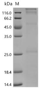
Recombinant Human Clarin-1 (CLRN1) is produced using an E.coli-based cell-free expression system and contains the full-length protein from amino acids 1 to 232. The protein has been engineered with an N-terminal 6xHis-tag to aid in purification, reaching a purity level greater than 85% as determined by SDS-PAGE. The protein features a four transmembrane domain structure and is presented using a detergent platform to maintain stability and functionality for research applications.

Clarin-1 appears to be a transmembrane protein that plays an important role in sensory perception, particularly within the auditory system. It seems to be involved in key cellular processes that may contribute to hearing function and maintenance. Understanding how Clarin-1 is structured and how it interacts with other components could be crucial for exploring its contributions to sensory pathways. This protein might also have potential implications in auditory research, as researchers study its impact on cellular signaling and sensory transduction.

Potential Applications

Note: The applications listed below are based on what we know about this protein's biological functions, published research, and experience from experts in the field. However, we haven't fully tested all of these applications ourselves yet. We'd recommend running some preliminary tests first to make sure they work for your specific research goals.

Based on the provided information, the recombinant Human CLRN1 is expressed using an in vitro E. coli expression system (cell-free system), which typically provides better folding conditions for membrane proteins by reducing cellular stress and aggregation. CLRN1 is a transmembrane protein with 4 transmembrane domains, making its proper folding challenging. The protein is full-length (1-232aa) with an N-terminal 6xHis tag, and purity is >85% by SDS-PAGE. However, since activity is unverified, the protein cannot be assumed to be correctly folded or bioactive. While the cell-free system improves folding probability, experimental validation is essential to confirm both proper folding (e.g., via circular dichroism or size exclusion chromatography) and bioactivity (e.g., through interaction assays with known binding partners).

1. Membrane Protein Biochemical Characterization Studies

This application is appropriate and should be prioritized. The recombinant CLRN1 can be used to study biochemical properties such as stability, detergent compatibility, and oligomerization state using techniques like size exclusion chromatography and dynamic light scattering. These studies can directly assess the protein's folding quality and are valuable even if the protein is not fully functional. The cell-free expression system provides a consistent protein preparation for reproducible characterization.

2. Antibody Development and Validation

This application is well-supported. The recombinant CLRN1 can serve as an effective immunogen for generating antibodies, as antibodies may recognize linear epitopes even if the protein is misfolded. The high purity (>85%) and His-tag facilitate purification and screening. However, if CLRN1 is misfolded, antibodies may not recognize conformational epitopes of the native protein. Validation against endogenous CLRN1 from mammalian cells is recommended to ensure physiological relevance.

3. Protein-Protein Interaction Studies

This application is feasible but requires caution. The His-tag enables pull-down assays to identify binding partners, but if CLRN1 is misfolded, interactions may be non-physiological. The 4-transmembrane domain structure is particularly sensitive to proper folding for native interactions. This application should only be pursued after confirming folding (e.g., via conformational antibodies or functional assays). Detergent solubilization may also alter some native interactions.

4. Membrane Protein Reconstitution Experiments

This application is high-risk without folding validation. While the detergent-solubilized CLRN1 can be reconstituted into lipid bilayers, if the protein is misfolded, its membrane integration behavior may not reflect native topology or function. Lipid composition studies may yield misleading data if the protein isn't properly structured. Folding should be confirmed before attempting reconstitution studies aimed at understanding biological mechanisms.

Final Recommendation & Action Plan

Given the uncertainty in folding and bioactivity, the recommended approach is to first perform comprehensive biochemical characterization (Application #1) to assess the protein's folding state, oligomerization, and stability. Techniques should include size exclusion chromatography (to check monodispersity), circular dichroism (for secondary structure), and thermal stability assays. If the protein shows proper folding, proceed with limited validation of protein-protein interactions using known binding partners. Antibody development (Application #2) can proceed concurrently, as it is less dependent on native conformation. Membrane reconstitution studies (Application #4) should be deferred until folding is validated. Always include appropriate controls, such as Western blotting with conformational antibodies when available, and compare results with literature on CLRN1 structure-function relationships where possible.

Customer Reviews and Q&A

 Customer Reviews

There are currently no reviews for this product.

Submit a Review here

Target Background

Function
May have a role in the excitatory ribbon synapse junctions between hair cells and cochlear ganglion cells and presumably also in analogous synapses within the retina.
Gene References into Functions
  1. Study founds 1 deep intronic CLRN1 (USH3A) founder mutation generates an aberrant exon that underlies severe Usher syndrome in a family on the Arabian Peninsula. PMID: 28469144
  2. This is the first report of Usher syndrome type 3 with a CLRN1 gene mutation in Asian populations. PMID: 25743179
  3. Possible digenism could not be excluded in two families segregating genomic variations in both MYO7A and USH2A, and two families with CLRN1 and USH2A. PMID: 22681893
  4. Two novel mutations in the CLRN1 gene, p.R207X and p.I168N, have been found in patients with Usher syndrome type 3. PMID: 23304067
  5. High-resolution measures of retinal structure demonstrate patterns of cone loss associated with CLRN1 mutations. PMID: 22964989
  6. This study confirmed using a novel mouse model carrying a Clrn1N48K knock-in mutation to investigate the consequence of the missense mutation N48K in mCLRN1 in vivo. PMID: 22787034
  7. Here we describe a novel deletion in CLRN1. Our data support previously reported intra familial variability in the clinical features of Usher syndrome type I and III. PMID: 21675857
  8. Retinitis pigmentosa-associated mutations p.Pro31Leu and p.Leu154Trp may represent hypomorphic mutations, because substituted amino acids in transmembrane domains remain polar. PMID: 21310491
  9. The complexity of the CLRN1 gene and the identification of multiple splice variants may partially explain why mutations in CLRN1 result in substantial variation in clinical phenotype. PMID: 20717163
  10. a role for clarin-1 in hair cell and photoreceptor cell synapses, as well as a common pathophysiological pathway for different Usher syndromes PMID: 12080385
  11. revised structure of USH3 gene: new translation start site, 5' untranslated region, and transcript encoding 232-amino acid protein; four new disease-causing mutations; identified mouse and rat orthologues, and two human paralogues on chromosomes 4 and 10 PMID: 12145752
  12. USH3A and USH2A share patterns of rod and cone dysfunction and retinal structural abnormalities. PMID: 18281613
  13. clarin-1 has a role in the regulation and homeostasis of actin filaments PMID: 19423712
  14. Part of the pathogenesis of USH3 may be associated with defective intracellular trafficking as well as decreased stability of mutant CLRN1 proteins. PMID: 19753315
  15. Mutations in the USH3 gene underlie Usher syndrome type 3. PMID: 11524702

Show More

Hide All

Involvement in disease
Usher syndrome 3A (USH3A); Retinitis pigmentosa 61 (RP61)
Subcellular Location
Cell membrane; Multi-pass membrane protein.
Protein Families
Clarin family
Tissue Specificity
Widely expressed. Found in the retina.
Database Links

HGNC: 12605

OMIM: 276902

KEGG: hsa:7401

UniGene: Hs.745448

icon of phone
Call us
301-363-4651 (Available 9 a.m. to 5 p.m. CST from Monday to Friday)
icon of address
Address
7505 Fannin St., Ste 610, Room 7 (CUBIO Innovation Center), Houston, TX 77054, USA
icon of social media
Join us with

Subscribe newsletter

Leave a message

* To protect against spam, please pass the CAPTCHA test below.
CAPTCHA verification
© 2007-2026 CUSABIO TECHNOLOGY LLC All rights reserved. 鄂ICP备15011166号-1
Place an order now

I. Product details

*
*
*
*

II. Contact details

*
*

III. Ship To

*
*
*
*
*
*
*

IV. Bill To

*
*
*
*
*
*
*
*