Call us
301-363-4651 (Available 9 a.m. to 5 p.m. CST from Monday to Friday)
| Code | CSB-CF407391HU |
| Abbreviation | Recombinant Human MYMK protein |
| MSDS | |
| Size | $1620 |
| Order now | |
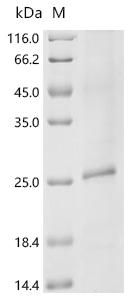
| Image |
|
| Have Questions? | Leave a Message or Start an on-line Chat |
Recombinant Human Protein myomaker (MYMK) is produced using an E.coli-based cell-free expression system, covering the full length of 1-221 amino acids. The protein features an N-terminal 10xHis-tag, which helps with purification and detection. It achieves a purity greater than 85% as determined by SDS-PAGE. The protein includes a 7-transmembrane domain and is presented using our proprietary Detergent Platform, ensuring optimal stability and functionality for research applications.
Myomaker appears to be a protein that's essential for muscle cell fusion—a process that seems crucial for muscle development and regeneration. It's involved in forming multinucleated muscle fibers and plays what may be a vital role in myogenesis. Studying this protein is likely important for understanding various muscle-related pathways and conditions, which makes it a significant focus in research on muscle biology and related disorders.
Potential Applications
Note: The applications listed below are based on what we know about this protein's biological functions, published research, and experience from experts in the field. However, we haven't fully tested all of these applications ourselves yet. We'd recommend running some preliminary tests first to make sure they work for your specific research goals.
The human MYMK is a multi-pass transmembrane protein with 7 transmembrane domains that requires proper integration into a lipid bilayer and correct folding for its bioactivity in mediating myoblast membrane fusion. While cell-free systems can improve solubility compared to traditional E. coli expression, they still lack the native membrane environment necessary for proper folding and integration of complex transmembrane proteins. The detergent used for solubilization may not replicate the natural lipid environment, potentially leading to non-native conformations. Without experimental validation (e.g., membrane integration assays or functional fusion assays), the protein cannot be assumed to be correctly folded or bioactive.
1. Membrane Protein Reconstitution Studies
This application is feasible but requires validation. The recombinant MYMK can be incorporated into artificial lipid bilayers to study membrane integration. However, if the protein is misfolded during expression, reconstitution may not yield functional protein. Validate membrane integration using techniques like fluorescence quenching or protease protection assays before concluding native behavior.
2. Protein-Protein Interaction Screening
Use with caution. The His-tag enables pull-down assays, but if MYMK is misfolded, interactions may be non-physiological. MYMK requires correct membrane integration and conformation for proper interactions with fusion partners. Validate any identified interactions using full-length MYMK expressed in mammalian cells or confirm with complementary methods like co-immunoprecipitation from myoblasts.
3. Antibody Development and Validation
This application is suitable. The recombinant MYMK can serve as an immunogen for generating antibodies against linear epitopes, even if misfolded. The high purity supports consistent immunization. However, antibodies generated may not recognize conformational epitopes of native, membrane-embedded MYMK. Validate antibody specificity against MYMK expressed in mammalian cells or endogenous MYMK in myoblasts.
4. Membrane Protein Biochemical Characterization
This recombinant human MYMK is suitable for basic biophysical analysis of the detergent-solubilized protein (e.g., thermal stability, aggregation state). However, data may not reflect native MYMK properties due to potential misfolding and detergent effects. Use techniques like circular dichroism to assess secondary structure, but interpret results cautiously without validation of membrane integration capability.
Final Recommendation & Action Plan
Before using this recombinant MYMK for functional studies, validate its folding and membrane integration capability. First, analyze secondary structure via circular dichroism and check oligomeric state via size-exclusion chromatography. Most critically, test membrane integration by reconstituting into proteoliposomes and assessing topology using protease protection assays. If possible, perform functional assays by co-expressing with MyoMixer in heterologous cells to test fusion activity. If active, proceed with interaction studies; if inactive, limit use to non-functional applications like antibody production (with validation against native MYMK). For reliable results, consider expressing MYMK in mammalian cells that provide proper membrane environments. Always include appropriate controls with native MYMK when possible.
There are currently no reviews for this product.

