| Image |
-
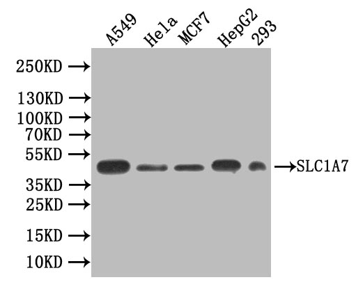
Western Blot
Positive WB detected in: A549 whole cell lysate, Hela whole cell lysate, MCF7 whole cell lysate, HepG2 whole cell lysate, 293 whole cell lysate
All lanes: SLC1A7 antibody at 1:1000
Secondary
Goat polyclonal to rabbit IgG at 1/50000 dilution
Predicted band size: 61 KD, 18 KD, 48 KD, 52 KD, 67 KD
Observed band size: 48 kDa
-
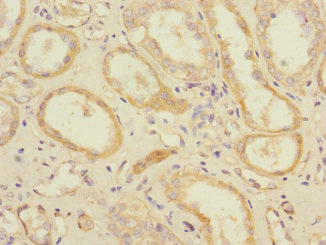
Immunohistochemistry of paraffin-embedded human kidney tissue using CSB-PA021438ESR2HU at dilution of 1:100
-
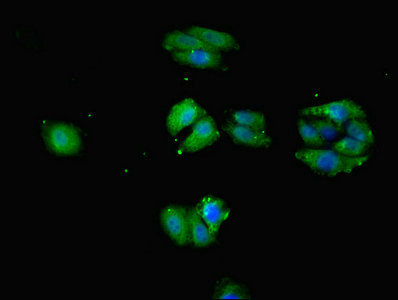
Immunofluorescent analysis of HepG2 cells using CSB-PA021438ESR2HU at dilution of 1:100 and Alexa Fluor 488-congugated AffiniPure Goat Anti-Rabbit IgG(H+L)
-
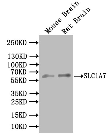
Western Blot
Positive WB detected in:Mouse Brain tissue lysate, Rat Brain tissue lysate
All lanes: SLC1A7 antibody at 1:1000
Secondary
Goat polyclonal to rabbit IgG at 1/50000 dilution
Predicted band size: 61 KD, 18 KD, 48 KD, 52 KD, 67 KD
Observed band size: 61 kDa
|