AGPAT5 Antibody

Code CSB-PA001453LA01HU
Size $297
Order now
Image
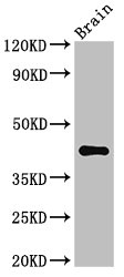
  • Western Blot
    Positive WB detected in: B16F10 whole cell lysate(30µg), A2780 whole cell lysate(30µg), Hela whole cell lysate(30µg), HL-60 whole cell lysate(30µg), THP-1 whole cell lysate(30µg), K562 whole cell lysate(20µg),SY5Y whole cell lysate(30µg),293 whole cell lysate(30µg),Mouse Brain tissue lysate(20µg)
    All lanes: AGPAT5 antibody at 1:1000
    Secondary
    Goat polyclonal to rabbit IgG at 1/40000 dilution
    Predicted band size: 43 kDa
    Observed band size: 43 kDa
    Exposure time: 60s
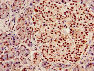
  • IHC image of CSB-PA001453LA01HU diluted at 1:100 and staining in paraffin-embedded human pancreatic tissue performed on a Leica BondTM system. After dewaxing and hydration, antigen retrieval was mediated by high pressure in a citrate buffer (pH 6.0). Section was blocked with 10% normal goat serum 30min at RT. Then primary antibody (1% BSA) was incubated at 4°C overnight. The primary is detected by a Goat anti-rabbit polymer IgG labeled by HRP and visualized using 0.05% DAB. Secondary antibody only control: uses 1% BSA instead of primary antibody
  • IHC image of CSB-PA001453LA01HU diluted at 1:100 and staining in paraffin-embedded human Liver cancer performed on a Leica BondTM system. After dewaxing and hydration, antigen retrieval was mediated by high pressure in a citrate buffer (pH 6.0). Section was blocked with 10% normal goat serum 30min at RT. Then primary antibody (1% BSA) was incubated at 4°C overnight. The primary is detected by a Goat anti-rabbit polymer IgG labeled by HRP and visualized using 0.05% DAB. Secondary antibody only control: uses 1% BSA instead of primary antibody
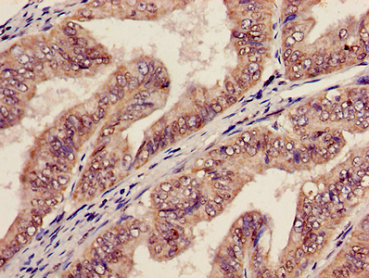
  • IHC image of CSB-PA001453LA01HU diluted at 1:100 and staining in paraffin-embedded human endometrial cancer performed on a Leica BondTM system. After dewaxing and hydration, antigen retrieval was mediated by high pressure in a citrate buffer (pH 6.0). Section was blocked with 10% normal goat serum 30min at RT. Then primary antibody (1% BSA) was incubated at 4°C overnight. The primary is detected by a Goat anti-rabbit polymer IgG labeled by HRP and visualized using 0.05% DAB. Secondary antibody only control: uses 1% BSA instead of primary antibody
Promotion Free Antibody trial simple
Have Questions? Leave a Message or Start an on-line Chat

Product Details

Full Product Name
Rabbit anti-Homo sapiens (Human) AGPAT5 Polyclonal antibody
Uniprot No.
Target Names
Alternative Names
AGPAT5; 1-acyl-sn-glycerol-3-phosphate acyltransferase epsilon; 1-acylglycerol-3-phosphate O-acyltransferase 5; 1-AGP acyltransferase 5; 1-AGPAT 5; Lysophosphatidic acid acyltransferase epsilon; LPAAT-epsilon
Raised in
Rabbit
Species Reactivity
Human, Mouse
Immunogen
Synthesized peptide derived from internal of human AGPAT5
Immunogen Species
Homo sapiens (Human)
Clonality
Polyclonal
Isotype
IgG
Purification Method
Antigen affinity purification
Concentration
It differs from different batches. Please contact us to confirm it.
Buffer
Rabbit IgG in phosphate buffered saline (without Mg2+ and Ca2+), pH 7.4, 150mM NaCl, 0.02% sodium azide and 50% glycerol
Form
Liquid
Tested Applications
ELISA, WB, IHC
Recommended Dilution
Application Recommended Dilution
WB 1:500-1:2000
IHC 1:20-1:200
Troubleshooting and FAQs
Storage
Upon receipt, store at -20°C or -80°C. Avoid repeated freeze.
Lead Time
Basically, we can dispatch the products out in 1-3 working days after receiving your orders. Delivery time maybe differs from different purchasing way or location, please kindly consult your local distributors for specific delivery time.
Usage
For Research Use Only. Not for use in diagnostic or therapeutic procedures.
Customer Reviews and Q&A

 Customer Reviews

There are currently no reviews for this product.

Submit a Review here

Target Background

Function
Converts 1-acyl-sn-glycerol-3-phosphate (lysophosphatidic acid or LPA) into 1,2-diacyl-sn-glycerol-3-phosphate (phosphatidic acid or PA) by incorporating an acyl moiety at the sn-2 position of the glycerol backbone. Acts on LPA containing saturated or unsaturated fatty acids C15:0-C20:4 at the sn-1 position using C18:1-CoA as the acyl donor. Also acts on lysophosphatidylethanolamine using oleoyl-CoA, but not arachidonoyl-CoA, and lysophosphatidylinositol using arachidonoyl-CoA, but not oleoyl-CoA. Activity toward lysophosphatidylglycerol not detectable.
Gene References into Functions
  1. enzymatic properties, tissue distribution, and subcellular localization of human AGPAT3 and AGPAT5 PMID: 21173190
  2. This publication describes the molecular cloning, tissue distribution, and enzyme characterization of the mouse homolog, mAGPAT5. PMID: 15367102
Subcellular Location
Endoplasmic reticulum membrane; Multi-pass membrane protein. Nucleus envelope. Mitochondrion.
Protein Families
1-acyl-sn-glycerol-3-phosphate acyltransferase family
Tissue Specificity
Widely expressed.
Database Links

HGNC: 20886

OMIM: 614796

KEGG: hsa:55326

STRING: 9606.ENSP00000285518

UniGene: Hs.624002

icon of phone
Call us
301-363-4651 (Available 9 a.m. to 5 p.m. CST from Monday to Friday)
icon of address
Address
7505 Fannin St., Ste 610, Room 7 (CUBIO Innovation Center), Houston, TX 77054, USA
icon of social media
Join us with

Subscribe newsletter

Leave a message

* To protect against spam, please pass the CAPTCHA test below.
CAPTCHA verification
© 2007-2026 CUSABIO TECHNOLOGY LLC All rights reserved. 鄂ICP备15011166号-1
Place an order now

I. Product details

*
*
*
*

II. Contact details

*
*

III. Ship To

*
*
*
*
*
*
*

IV. Bill To

*
*
*
*
*
*
*
*