
Call us
301-363-4651 (Available 9 a.m. to 5 p.m. CST from Monday to Friday)
| Code | CSB-PA851843HA01MO |
| Size | US$166 |
| Order now | |
| Image |
|
| Promotion |  |
| Have Questions? | Leave a Message or Start an on-line Chat |
The Plet1 Antibody (Product code: CSB-PA851843HA01MO) is Non-conjugated. For Plet1 Antibody with conjugates, please check the following table.
| Application | Recommended Dilution |
|---|---|
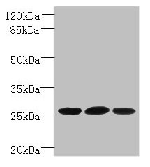
| WB | 1:500-1:2000 |
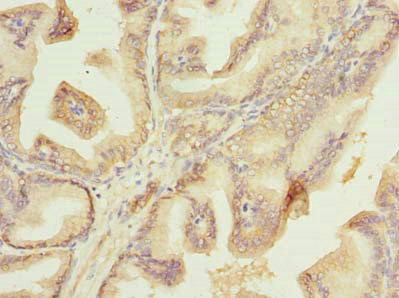
| IHC | 1:20-1:200 |
To generate the Plet1 polyclonal antibody, the recombinant mouse Plet1 protein (28-218aa) was administered to a rabbit to induce an immune response. The rabbit blood was collected and purified using protein G affinity chromatography, getting the Plet1 polyclonal antibody. The purity of the Plet1 antibody obtained through this process is over 95%, which ensures the reliability and accuracy of the results.
This high-purity Plet1 antibody has demonstrated reactivity with both human and mouse samples and has been thoroughly validated for use in various applications, including ELISA, WB, and IHC. These applications enable the detection of the presence and quantity of Plet1 protein, the visualization of its distribution and localization, as well as its identification.
There are currently no reviews for this product.

