WIPI1 Antibody

Code CSB-PA705810ESR2HU
Size US$166
Order now
Image
  • Western blot
    All lanes: WIPI1 antibody at 6μg/ml
    Lane 1: 293T whole cell lysate
    Lane 2: NIH/3T3 whole cell lysate
    Lane 3: Mouse skeletal muscle tissue
    Secondary
    Goat polyclonal to rabbit IgG at 1/10000 dilution
    Predicted band size: 49 kDa
    Observed band size: 49 kDa

  • Immunohistochemistry of paraffin-embedded human placenta tissue using CSB-PA705810ESR2HU at dilution of 1:100

Have Questions? Leave a Message or Start an on-line Chat

Product Details

Full Product Name
Rabbit anti-Homo sapiens (Human) WIPI1 Polyclonal antibody
Uniprot No.
Target Names
WIPI1
Alternative Names
ATG 18 antibody; ATG18 antibody; Atg18 protein homolog antibody; ATG18A antibody; FLJ10055 antibody; WD repeat domain phosphoinositide interacting 1 antibody; WD repeat domain phosphoinositide interacting protein 1 antibody; WD repeat domain phosphoinositide-interacting protein 1 antibody; WD40 repeat protein interacting with phosphoinositides of 49 kDa antibody; WD40 repeat protein interacting with phosphoInositides of 49kDa antibody; WIPI 1 alpha antibody; WIPI 1 antibody; WIPI 49 antibody; WIPI 49 kDa antibody; WIPI-1 antibody; wipi1 antibody; WIPI1_HUMAN antibody; WIPI49 antibody
Raised in
Rabbit
Species Reactivity
Human, Mouse
Immunogen
Recombinant Human WD repeat domain phosphoinositide-interacting protein 1 protein (187-446AA)
Immunogen Species
Homo sapiens (Human)
Conjugate
Non-conjugated
Clonality
Polyclonal
Isotype
IgG
Purification Method
Antigen Affinity Purified
Concentration
It differs from different batches. Please contact us to confirm it.
Buffer
PBS with 0.02% sodium azide, 50% glycerol, pH7.3.
Form
Liquid
Tested Applications
ELISA, WB, IHC
Recommended Dilution
Application Recommended Dilution
WB 1:500-1:2000
IHC 1:20-1:200
Troubleshooting and FAQs
Storage
Upon receipt, store at -20°C or -80°C. Avoid repeated freeze.
Lead Time
Basically, we can dispatch the products out in 1-3 working days after receiving your orders. Delivery time maybe differs from different purchasing way or location, please kindly consult your local distributors for specific delivery time.

Customer Reviews and Q&A

 Customer Reviews

There are currently no reviews for this product.

Submit a Review here

Target Background

Function
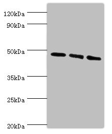
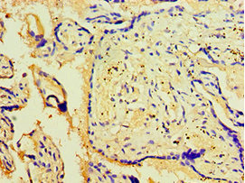
Component of the autophagy machinery that controls the major intracellular degradation process by which cytoplasmic materials are packaged into autophagosomes and delivered to lysosomes for degradation. Plays an important role in starvation- and calcium-mediated autophagy, as well as in mitophagy. Functions downstream of the ULK1 and PI3-kinases that produce phosphatidylinositol 3-phosphate (PtdIns3P) on membranes of the endoplasmic reticulum once activated. Binds phosphatidylinositol 3-phosphate (PtdIns3P), and maybe other phosphoinositides including PtdIns3,5P2 and PtdIns5P, and is recruited to phagophore assembly sites at the endoplasmic reticulum membranes. There, it assists WIPI2 in the recruitment of ATG12-ATG5-ATG16L1, a complex that directly controls the elongation of the nascent autophagosomal membrane. Together with WDR45/WIPI4, promotes ATG2 (ATG2A or ATG2B)-mediated lipid transfer by enhancing ATG2-association with phosphatidylinositol 3-monophosphate (PI3P)-containing membranes. Involved in xenophagy of Staphylococcus aureus. Invading S.aureus cells become entrapped in autophagosome-like WIPI1 positive vesicles targeted for lysosomal degradation. Plays also a distinct role in controlling the transcription of melanogenic enzymes and melanosome maturation, a process that is distinct from starvation-induced autophagy. May also regulate the trafficking of proteins involved in the mannose-6-phosphate receptor (MPR) recycling pathway.
Gene References into Functions
  1. The specific autophagosomal localization of both WIPI1 and WIPI2 (refered to as WIPI puncta) has been employed to assess autophagy using fluorescence microscopy methods, such as confocal and live-cell video microscopy PMID: 25462558
  2. Data suggest WIPI1/WIPI2 co-localize with microtubule-associated light chain 3 and autophagy related proteins 2/14L, participate in biogenesis of phagosomes, autophagy, and mobilization of lipids to/from intracellular droplets. [review-like article] PMID: 25233424
  3. WIPI-1 and WIPI-2 are functionally required in mediating the PI3P signal at the onset of autophagy in NB4 cells. PMID: 24991767
  4. the detection of WIPI1 mRNA is likely to be a convenient method of monitoring autophagosome formation in a wide range of cell types PMID: 24384561
  5. Freeze-fracture replica immunolabelling reveals WD-repeat protein interacting with phosphoinositides 1 and 2 (WIPI-1 and WIPI-2) as membrane components of autophagosomes and the plasma membrane (PM). PMID: 21564513
  6. Studies define a distinct role for WIPI1 and TORC1 signaling in controlling the transcription of melanogenic enzymes and melanosome maturation, a process that is distinct from starvation-induced autophagy. PMID: 21317285
  7. share an evolutionary conserved function in autophagy and that autophagic capacity may be compromised in human cancers PMID: 15602573
  8. Quantification of WIPI-1 puncta should be suitable to assay mammalian autophagy PMID: 17618624

Show More

Hide All

Subcellular Location
Golgi apparatus, trans-Golgi network. Endosome. Cytoplasmic vesicle, clathrin-coated vesicle. Preautophagosomal structure membrane; Peripheral membrane protein. Cytoplasm, cytoskeleton. Note=Trans elements of the Golgi and peripheral endosomes. Dynamically cycles through these compartments and is susceptible to conditions that modulate membrane flux. Enriched in clathrin-coated vesicles. Upon starvation-induced autophagy, accumulates at subcellular structures in the cytoplasm: enlarged vesicular and lasso-like structures, and large cup-shaped structures predominantly around the nucleus. Recruitment to autophagic membranes is controlled by MTMR14. Labile microtubules specifically recruit markers of autophagosome formation like WIPI1, whereas mature autophagosomes may bind to stable microtubules.
Protein Families
WD repeat SVP1 family
Tissue Specificity
Ubiquitously expressed. Highly expressed in skeletal muscle, heart, testis, pancreas and placenta. Highly expressed in G361, Sk-mel-28, Sk-mel-13, WM852 and WM451 cells. Up-regulated in a variety of tumor tissues.
Database Links

HGNC: 25471

OMIM: 609224

KEGG: hsa:55062

STRING: 9606.ENSP00000262139

UniGene: Hs.463964

icon of phone
Call us
301-363-4651 (Available 9 a.m. to 5 p.m. CST from Monday to Friday)
icon of address
Address
7505 Fannin St., Ste 610, Room 7 (CUBIO Innovation Center), Houston, TX 77054, USA
icon of social media
Join us with

Subscribe newsletter

Leave a message

* To protect against spam, please pass the CAPTCHA test below.
CAPTCHA verification
© 2007-2024 CUSABIO TECHNOLOGY LLC All rights reserved. 鄂ICP备15011166号-1
Place an order now

I. Product details

*
*
*
*

II. Contact details

*
*

III. Ship To

*
*
*
*
*
*
*

IV. Bill To

*
*
*
*
*
*
*
*