Call us
301-363-4651 (Available 9 a.m. to 5 p.m. CST from Monday to Friday)
| Code | CSB-EP852881HU |
| Abbreviation | Recombinant Human PIEZO1 protein, partial |
| MSDS | |
| Size | $306 |
| Order now | |
| Image |
|
| Have Questions? | Leave a Message or Start an on-line Chat |
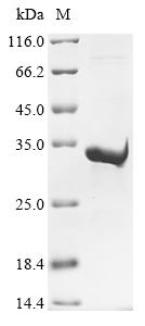
The recombinant human PIEZO1 protein is labeled with an N-terminal 10xHis-tagged and C-terminal Myc-tag. It is expressed in the E.coli. The expression region corresponds to the 2198-2431aa of the human PIEZO1. It is subject to affinity chromatography purification. Its purity is over 90% as measured by SDS-PAGE.
Human PIEZO1 functions as a mechanosensitive ion channel and is primarily involved in transducing mechanical stimuli into cellular responses. Upon stimulation by mechanical forces, such as membrane stretch or deformation, PIEZO1 occurs a conformational change, allowing ion influx [1-3]. This mechanotransduction process is vital to regulating calcium signaling, which is crucial for muscle contraction, neurotransmitter release, and cellular signaling pathways [4]. It is a non-selective cation channel that allows the passage of various ions, including Na+, K+, and Ca2+, thereby playing a significant role in various physiological processes such as cell proliferation, differentiation, and apoptosis [5][6].
Research has demonstrated that PIEZO1 is involved in several pathophysiological conditions. The dysregulation of PIEZO1 has been linked to diseases such as multiple sclerosis, where its expression is downregulated in the brain, affecting oligodendrocyte maturation and migration [7]. PIEZO1 has been implicated in cancer biology, influencing tumor progression and immune responses by modulating calcium signaling in immune cells [4].
References:
[1] J. Roh, S. Hwang, S. Lee, K. Lee, Y. Kim, & C. Park, Functional expression of piezo1 in dorsal root ganglion (drg) neurons, International Journal of Molecular Sciences, vol. 21, no. 11, p. 3834, 2020. https://doi.org/10.3390/ijms21113834
[2] M. Yao, A. Tijore, D. Cheng, J. Li, A. Hariharan, B. Martinacet al., Force- and cell state–dependent recruitment of piezo1 drives focal adhesion dynamics and calcium entry, Science Advances, vol. 8, no. 45, 2022. https://doi.org/10.1126/sciadv.abo1461
[3] A. Schröder, K. Neher, B. Krenmayr, E. Paddenberg, G. Spanier, P. Proffet al., Impact of piezo1‐channel on inflammation and osteoclastogenesis mediated via periodontal ligament fibroblasts during mechanical loading, European Journal of Oral Sciences, vol. 131, no. 1, 2023. https://doi.org/10.1111/eos.12913
[4] Y. Wu, J. Zhang, C. Hou, H. Wang, M. Zhu, & X. Yao, A pancancer study of piezo1 as a prognosis and immune biomarker of human tumors, Journal of Oncology, vol. 2022, p. 1-15, 2022. https://doi.org/10.1155/2022/6725570
[5] B. Wang, W. Ke, K. Wang, G. Li, L. Ma, S. Luet al., Mechanosensitive ion channel piezo1 activated by matrix stiffness regulates oxidative stress‐induced senescence and apoptosis in human intervertebral disc degeneration, Oxidative Medicine and Cellular Longevity, vol. 2021, no. 1, 2021. https://doi.org/10.1155/2021/8884922
[6] X. Ren, H. Zhuang, B. Li, F. Jiang, Y. Zhang, & P. Zhou, Gsmtx4 alleviated osteoarthritis through piezo1/calcineurin/nfat1 signaling axis under excessive mechanical strain, International Journal of Molecular Sciences, vol. 24, no. 4, p. 4022, 2023. https://doi.org/10.3390/ijms24044022
[7] M. Velasco‐Estevez, N. Koch, I. Klejbor, F. Caratis, & A. Rutkowska, Mechanoreceptor piezo1 is downregulated in multiple sclerosis brain and is involved in the maturation and migration of oligodendrocytes in vitro, Frontiers in Cellular Neuroscience, vol. 16, 2022. https://doi.org/10.3389/fncel.2022.914985
There are currently no reviews for this product.

