Recombinant Mouse Cystatin-C (Cst3)

In Stock
Code CSB-EP006091MO
Abbreviation Recombinant Mouse Cst3 protein
MSDS
Size $388
Order now
Image
  • (Tris-Glycine gel) Discontinuous SDS-PAGE (reduced) with 5% enrichment gel and 15% separation gel.
Have Questions? Leave a Message or Start an on-line Chat

Product Details

Purity
Greater than 85% as determined by SDS-PAGE.
Target Names
Uniprot No.
Research Area
Cardiovascular
Alternative Names
Cst3Cystatin-C; Cystatin-3
Species
Mus musculus (Mouse)
Source
E.coli
Expression Region
21-140aa
Target Protein Sequence
ATPKQGPRMLGAPEEADANEEGVRRALDFAVSEYNKGSNDAYHSRAIQVVRARKQLVAGVNYFLDVEMGRTTCTKSQTNLTDCPFHDQPHLMRKALCSFQIYSVPWKGTHSLTKFSCKNA
Note: The complete sequence may include tag sequence, target protein sequence, linker sequence and extra sequence that is translated with the protein sequence for the purpose(s) of secretion, stability, solubility, etc.
If the exact amino acid sequence of this recombinant protein is critical to your application, please explicitly request the full and complete sequence of this protein before ordering.
Mol. Weight
20.4 kDa
Protein Length
Full Length of Mature Protein
Tag Info
N-terminal 10xHis-tagged and C-terminal Myc-tagged
Form
Liquid or Lyophilized powder
Note: We will preferentially ship the format that we have in stock, however, if you have any special requirement for the format, please remark your requirement when placing the order, we will prepare according to your demand.
Buffer
If the delivery form is liquid, the default storage buffer is Tris/PBS-based buffer, 5%-50% glycerol.
Note: If you have any special requirement for the glycerol content, please remark when you place the order.
If the delivery form is lyophilized powder, the buffer before lyophilization is Tris/PBS-based buffer, 6% Trehalose.
Reconstitution
We recommend that this vial be briefly centrifuged prior to opening to bring the contents to the bottom. Please reconstitute protein in deionized sterile water to a concentration of 0.1-1.0 mg/mL.We recommend to add 5-50% of glycerol (final concentration) and aliquot for long-term storage at -20°C/-80°C. Our default final concentration of glycerol is 50%. Customers could use it as reference.
Troubleshooting and FAQs
Storage Condition
Store at -20°C/-80°C upon receipt, aliquoting is necessary for mutiple use. Avoid repeated freeze-thaw cycles.
Shelf Life
The shelf life is related to many factors, storage state, buffer ingredients, storage temperature and the stability of the protein itself.
Generally, the shelf life of liquid form is 6 months at -20°C/-80°C. The shelf life of lyophilized form is 12 months at -20°C/-80°C.
Lead Time
3-7 business days
Notes
Repeated freezing and thawing is not recommended. Store working aliquots at 4°C for up to one week.
Datasheet & COA
Please contact us to get it.
Description

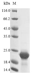
Recombinant Mouse Cystatin-C (Cst3) is produced in E. coli and includes an N-terminal 10xHis-tag along with a C-terminal Myc-tag, which makes purification and detection more straightforward. The protein represents the complete mature form, covering amino acids 21-140. SDS-PAGE analysis confirms purity levels above 85%, making this product well-suited for research applications that demand highly specific and consistent recombinant protein.

Cystatin-C appears to be a cysteine protease inhibitor that researchers study extensively for its regulatory function in proteolytic pathways. It seems crucial for maintaining protease balance and may have important implications in research focusing on tissue remodeling, inflammation, and cellular homeostasis. Understanding how it interacts with other molecules is likely essential for revealing the mechanisms behind protease-related processes in biological systems.

Potential Applications

Note: The applications listed below are based on what we know about this protein's biological functions, published research, and experience from experts in the field. However, we haven't fully tested all of these applications ourselves yet. We'd recommend running some preliminary tests first to make sure they work for your specific research goals.

Mouse Cst3 is a cysteine protease inhibitor that requires precise folding, proper disulfide bond formation (with two conserved disulfide bonds), and specific tertiary structure for its functional activity in cathepsin inhibition. Cystatin-C is a small-molecular-weight extracellular inhibitor protein. Its activity does not rely on any complex post-translational modifications. Its function depends on forming the correct three-dimensional structure, which includes a pocket for binding to the target enzyme (cysteine protease). This structure is relatively stable and is relatively easy to fold correctly in the cytoplasmic environment of Escherichia coli. The dual N-terminal 10xHis-tag and C-terminal Myc-tag may sterically interfere with the protein's functional domains, particularly the protease-binding sites. While the protein may be soluble, the probability of correct folding with functional protease inhibitory activity is low without experimental validation.

1. Cysteine Protease Inhibition Assays

This application carries a significant risk without proper folding validation. Cystatin-C's inhibitory function requires precise tertiary structure and proper disulfide bonding for cathepsin binding. If correctly folded and active (verified through inhibition assays), the protein may be suitable for kinetic studies. If misfolded/inactive (unverified), inhibition assays will yield biologically meaningless results. The dual tags may sterically block the protease-binding interface.

2. Antibody Development and Validation

This application is highly suitable as antibody development relies on antigenic sequence recognition rather than functional protein folding. The full-length mature protein provides comprehensive epitope coverage for generating antibodies against mouse Cystatin-C. The dual tags offer additional epitopes for screening and validation purposes.

3. Protein-Protein Interaction Studies

This application requires proper folding validation. Cystatin-C interactions with proteases require precise tertiary structure. If correctly folded (verified), the protein may identify physiological interaction partners. If misfolded/unverified, there is a high risk of non-specific binding or failure to replicate genuine protease-inhibitor interactions.

4. Structural and Biophysical Characterization

These studies are essential for determining the folding status of the protein itself, not the native Cst3. Techniques should include circular dichroism spectroscopy to assess secondary structure, size-exclusion chromatography to evaluate oligomeric state, and disulfide bond analysis. However, the dual tags may interfere with crystallization for high-resolution structural studies.

5. Comparative Species Studies

Meaningful comparative studies require native protein conformation and functional activity. If correctly folded and active (verified), the protein enables valid evolutionary comparisons of inhibitory properties across species. If misfolded/inactive (unverified), comparative analyses would yield misleading evolutionary insights

Final Recommendation & Action Plan

The E. coli expression system is fundamentally limited for producing functional Cystatin-C due to its inability to support proper disulfide bond formation. Begin with Application 4 (Structural Characterization) to assess folding quality through CD spectroscopy, SEC, and validate disulfide bond formation. Test inhibitory activity against known cathepsins before considering functional applications. Applications 1, 3, and 5 require rigorous functional validation. Application 2 (antibody development) can proceed immediately. For reliable Cystatin-C research requiring native functionality, use mammalian expression systems that support proper disulfide bonding and folding.

Customer Reviews and Q&A

 Customer Reviews

There are currently no reviews for this product.

Submit a Review here

Target Background

Function
As an inhibitor of cysteine proteinases, this protein is thought to serve an important physiological role as a local regulator of this enzyme activity.
Gene References into Functions
  1. CysC plays an important regulatory role in combating cell death via the autophagic pathway in atherosclerosis. PMID: 27079462
  2. Data (including data from studies in mutant mice) suggest that up-regulation of Cst3, as observed in plasma of mice with type 2 diabetes, down-regulates insulin signaling and promotes endoplasmic reticulum stress in hepatocytes but not in myotubes. PMID: 26592151
  3. Cystatin C is a potential pathogenic signal triggering neurodegeneration in multiple system atrophy. PMID: 24405769
  4. The neuroprotective activity of CysC against Amyotrophic lateral sclerosis-linked mutant Cu/Zn-superoxide dismutase (SOD1)-mediated toxicity. PMID: 25356866
  5. an important role for macrophages, DC, and ROS in diseases associated with the protease inhibitory activity or amyloidogenic properties of cystatin C. PMID: 24570004
  6. APP expression stimulates NSPC proliferation; this effect is mediated via an increase in cystatin C secretion PMID: 23671283
  7. Cystatin C, a protein targeted to the classical secretory pathway by its signal peptide sequence, is secreted by primary neurons in at least 9 different cystatin C glycoforms associated with exosomes. PMID: 19773092
  8. The lack of cystatin C enhances Collagen-induced arthritis and primarily affects in vivo priming of the immune system PMID: 21443774
  9. Inflammation causes downregulation of cystatin C expression in dendritic cells and reduces serum CstC levels. PMID: 21300820
  10. Data show that the absence of cystatin C reduced epithelial cell apoptosis but increased proliferation in skin. PMID: 21085595
  11. Data show that cystatin C effectively rescues cystatin B loss-of-function mutation, facilitating the reversal of pathophysiological changes and suggesting a novel therapeutic intervention for patients with neurodegenerative disorders. PMID: 20889561
  12. CysC plays a protective role under conditions of neuronal challenge by inducing autophagy via mTOR inhibition PMID: 20352108
  13. Oxidants induce elevated cystatin C production from cardiac myocytes. Cystatin C plays a role in cardiac extracellular matrix remodelling. PMID: 20489058
  14. Data show that cystatin c (CystC) contributes to experimental abdominal aortic aneurysms (AAA) pathogenesis and that lack of CystC favors inflammation in AAA lesions induced in atherosclerotic mice. PMID: 20472891
  15. The embryo's cystatin C and F expression functions as a protective mechanism against the maternal proteinase cathepsin S. PMID: 20093401
  16. Cystatin C through this effect can act as an immunomodulatory molecule. PMID: 19446036
  17. expressed in decidual cultures and the expression is controlled in part by TGF-beta(1) and EGF signaling PMID: 11803549
  18. spatiotemporal up-regulation of cystatin C in the hippocampus is induced by entorhinal deafferentation (perforant path transections) PMID: 12044447
  19. Cystatin C is neither necessary nor sufficient to control expression of major histocompatibility type II complexes and antigen presentation in dendritic cells. PMID: 14607896
  20. In vivo overexpression of human cystatin C does not affect amyloid-beta levels in mice that do not deposit amyloid-beta. PMID: 14742906
  21. Data indicate that cystatin C is involved in astrocyte differentiation during mouse brain development. PMID: 15509901
  22. leukocyte-specific expression of cystatin C is actively involved in matrix remodelling associated with atherosclerosis plaque regression. PMID: 15823274
  23. the protective role of cystatin C in atherosclerosis is dependent primarily on its expression in nonhematopoietic cell types PMID: 16051881
  24. PKB activity is a critical downstream component of PDK-1 in mediating translation of cystatin C, RANKL, and Rab11a PMID: 16166629
  25. These data demonstrate that cystatin C has a role in neuronal death and neurogenesis during SE-induced network reorganization. PMID: 16242633
  26. Validity of cystatin C for generating neural stem cells from embryo stem cells in vitro. PMID: 16595632
  27. We propose that in MS, remyelination may be impaired by increasing activity of cathepsins inadequately controlled by cystatin C. PMID: 17086443
  28. Finally, in vivo studies using kidney-specific megalin knockout mice provide evidence that megalin mediates proximal tubular uptake of cystatin C. We conclude that megalin is an endocytic receptor of cystatin C in PTC. PMID: 17462596
  29. These results suggest that CysC affects the BMP signaling cascades in osteoblastic cells and then promotes osteoblast differentiation, mineralization, and bone formation. PMID: 17592728
  30. Plasminogen activator inhibitor-2, but not cystatin C, inhibits the prometastatic activity of tissue inhibitor of metalloproteinases-1 in the liver.( PMID: 18681831
  31. CysC regulates soluble Abeta and Abeta-associated neuronal deficits through inhibiting CatB-induced Abeta degradation. PMID: 18957217

Show More

Hide All

Subcellular Location
Secreted.
Protein Families
Cystatin family
Database Links
icon of phone
Call us
301-363-4651 (Available 9 a.m. to 5 p.m. CST from Monday to Friday)
icon of address
Address
7505 Fannin St., Ste 610, Room 7 (CUBIO Innovation Center), Houston, TX 77054, USA
icon of social media
Join us with

Subscribe newsletter

Leave a message

* To protect against spam, please pass the CAPTCHA test below.
CAPTCHA verification
© 2007-2025 CUSABIO TECHNOLOGY LLC All rights reserved. 鄂ICP备15011166号-1
×
Place an order now

I. Product details

*
*
*
*

II. Contact details

*
*

III. Ship To

*
*
*
*
*
*
*

IV. Bill To

*
*
*
*
*
*
*
*