Call us
301-363-4651 (Available 9 a.m. to 5 p.m. CST from Monday to Friday)
| Code | CSB-RA216446A0HU |
| Size | US$210 |
| Order now | |
| Image |
|
| Have Questions? | Leave a Message or Start an on-line Chat |
| Application | Recommended Dilution |
|---|---|
| WB | 1:500-1:5000 |
| IHC | 1:50-1:200 |
ATP5F1B, the beta subunit of mitochondrial ATP synthase, serves as the catalytic core of the enzyme complex responsible for oxidative phosphorylation—the fundamental process by which cells generate ATP. This protein plays a central role in cellular energy metabolism, making it an essential marker for studies investigating mitochondrial function, metabolic reprogramming in cancer, and bioenergetic pathways across diverse tissue types.
This recombinant monoclonal antibody, generated from clone 5F10, offers the reproducibility and consistency that demanding experimental workflows require. Because recombinant antibodies are produced from defined sequences rather than traditional hybridoma methods, researchers benefit from lot-to-lot uniformity that supports longitudinal studies and ensures comparable results across experiments. The rabbit IgG format provides excellent signal-to-noise characteristics for detection applications.
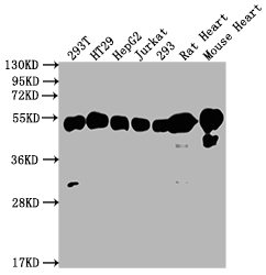
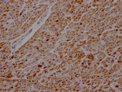
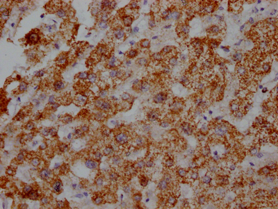
Validation data demonstrates robust performance across multiple platforms. In western blot applications, this antibody detects a clean band at the expected 57 kDa molecular weight across a range of human cell lines including 293T, HT29, HepG2, Jurkat, and 293 cells, with effective dilutions ranging from 1:500 to 1:5000. Cross-species reactivity extends to rodent samples, with confirmed detection in both rat and mouse heart tissue, providing flexibility for researchers working with animal models. Immunohistochemistry validation in paraffin-embedded human heart and liver tissue at 1:100 dilution shows specific staining patterns consistent with the mitochondria-rich nature of these metabolically active organs.
Whether investigating tumor metabolism, characterizing mitochondrial dysfunction, or exploring signal transduction pathways linked to cellular energetics, this antibody provides a reliable tool for detecting ATP5F1B across your experimental systems.
There are currently no reviews for this product.

