ATP6V1A Recombinant Monoclonal Antibody

Code CSB-RA193957A0HU
Size US$210
Order now
Image
  • Flow cytometric analysis of ATP6V1A expression in HepG2 cells using ATP6V1A antibody. Green, isotype control; red, ATP6V1A.
  • Immunocytochemical staining of HepG2 cells with ATP6V1A antibody. Nuclei were stained blue with DAPI; ATP6V1A was stained magenta with Alexa Fluor® 647. Images were taken using Leica stellaris 5. Protein abundance based on laser Intensity and smart gain: Medium. Scale bar, 20 μm.
  • Immunohistochemistry was performed on paraffin-embedded human breast carcinoma using ATP6V1A antibody. Antigen retrieval was done in sodium citrate buffer (pH 6.0). DAB was used for detection, with hematoxylin counterstaining. Images were acquired using a Nikon Ci-L Plus microscope (40× objective). Scale bar: 25 μm.
  • Western blotting analysis using ATP6V1A antibody. Total cell lysates (30 μg) from various cell lines were loaded and separated by SDS-PAGE. The blot was incubated with ATP6V1A antibody and HRP-conjugated goat anti-rabbit secondary antibody respectively.
Have Questions? Leave a Message or Start an on-line Chat

Product Details

Uniprot No.
Target Names
Alternative Names
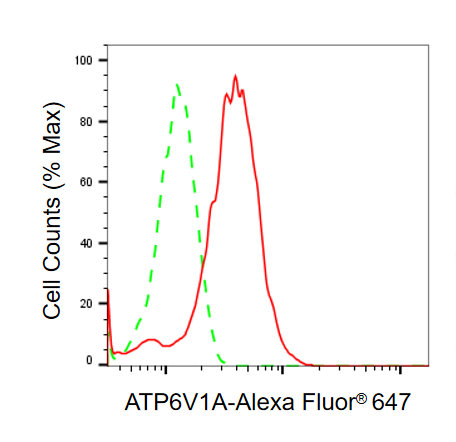
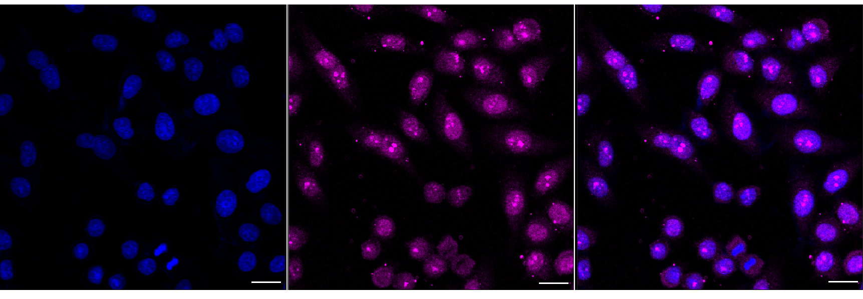
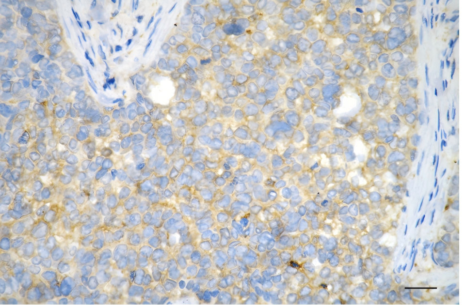
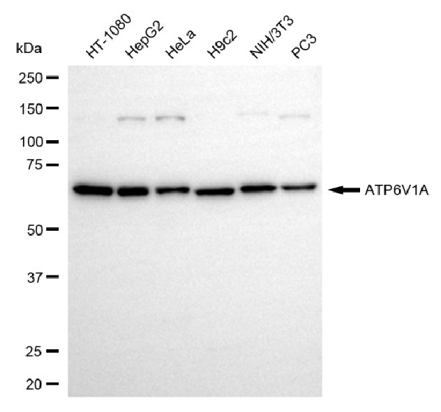
ATP6V1A; ATPase H+ Transporting V1 Subunit A; V-ATPase Subunit A; ATP6V1A1; ATP6A1; Vma1; VA68; VPP2; ATPase, H+ Transporting, Lysosomal 70kDa, V1 Subunit A; V-Type Proton ATPase (V-ATPase) Catalytic Subunit A; V-Type Proton ATPase Catalytic Subunit A; Vacuolar Proton Pump Subunit Alpha; ATPase, H+ Transporting, Lysosomal (Vacuolar Proton Pump), Alpha Polypeptide, 70kD, Isoform 1; H+-Transporting ATPase Chain A, Vacuolar (VA68 Type); ATPase, H+ Transporting, Lysosomal, Subunit A1; H(+)-Transporting Two-Sector ATPase, Subunit A; Vacuolar Proton Pump Alpha Subunit 1; Vacuolar ATPase Isoform VA68; V-ATPase 69 KDa Subunit 1; Vacuolar-Type H(+)-ATPase; V-ATPase 69 KDa Subunit; V-ATPase A Subunit 1; EC 3.6.3.14; EC 7.1.2.2; EC 3.6.3; ARCL2D; IECEE3; DEE93; HO68
Species Reactivity
Human, Mouse, Rat
Immunogen
Recombinant Human ATP6V1A protein
Immunogen Species
Homo sapiens (Human)
Conjugate
Non-conjugated
Clonality
Monoclonal
Isotype
Rabbit IgG
Clone No.
2D3
Purification Method
Affinity-chromatography
Concentration
It differs from different batches. Please contact us to confirm it.
Buffer
Rabbit IgG in PBS (pH 7.4) containing 50% glycerol, and 0.02% sodium azide.
Form
Liquid
Tested Applications
ELISA, WB, FC, ICC, IHC
Recommended Dilution
Application Recommended Dilution
WB 1:1000-1:5000
FC 1:200-1:2000
ICC 1:100-1:1000
IHC 1:100-1:200
Troubleshooting and FAQs
Storage
Upon receipt, store at -20°C or -80°C. Avoid repeated freeze.
Lead Time
Basically, we can dispatch the products out in 1-3 working days after receiving your orders. Delivery time maybe differs from different purchasing way or location, please kindly consult your local distributors for specific delivery time.
Usage
For Research Use Only. Not for use in diagnostic or therapeutic procedures.

Customer Reviews and Q&A

 Customer Reviews

There are currently no reviews for this product.

Submit a Review here

Target Background

Function
Catalytic subunit of the V1 complex of vacuolar(H+)-ATPase (V-ATPase), a multisubunit enzyme composed of a peripheral complex (V1) that hydrolyzes ATP and a membrane integral complex (V0) that translocates protons. V-ATPase is responsible for acidifying and maintaining the pH of intracellular compartments and in some cell types, is targeted to the plasma membrane, where it is responsible for acidifying the extracellular environment. In aerobic conditions, involved in intracellular iron homeostasis, thus triggering the activity of Fe(2+) prolyl hydroxylase (PHD) enzymes, and leading to HIF1A hydroxylation and subsequent proteasomal degradation. May play a role in neurite development and synaptic connectivity.; (Microbial infection) Plays an important role in virion uncoating during Rabies virus replication after membrane fusion. Specifically, participates in the dissociation of incoming viral matrix M proteins uncoating through direct interaction.
Gene References into Functions
  1. We report biallelic mutations in ATP6V1E1 and ATP6V1A, respectively encoding the E1 and A subunits of the V1 domain of V-ATPase, as a cause of distinct metabolic and multisystemic cutis laxa entities. PMID: 28065471
  2. V1A subunit of V-ATPase has a role in the progression and prognosis of gastric cancer. PMID: 25652905
  3. Data show that the cAMP/PKA/CREB signaling pathway initiates acidosis-induced V-ATPase trafficking in salivary ducts via regulation of Rab11b expression. PMID: 22561749
  4. These data introduce Rab11b as a crucial regulator and Rip11 as mediator of acidosis-induced V-ATPase traffic in duct cells of submandibular gland. PMID: 20717956
  5. there is an important role for physical association between aldolase and the A, B and E subunits of V-ATPase in the regulation of the proton pump PMID: 17576770

Show More

Hide All

Involvement in disease
Cutis laxa, autosomal recessive, 2D (ARCL2D)
Subcellular Location
Cytoplasm. Cytoplasmic vesicle, secretory vesicle.
Protein Families
ATPase alpha/beta chains family
Tissue Specificity
High expression in the skin.
Database Links

HGNC: 851

OMIM: 607027

KEGG: hsa:523

STRING: 9606.ENSP00000273398

UniGene: Hs.477155

icon of phone
Call us
301-363-4651 (Available 9 a.m. to 5 p.m. CST from Monday to Friday)
icon of address
Address
7505 Fannin St., Ste 610, Room 7 (CUBIO Innovation Center), Houston, TX 77054, USA
icon of social media
Join us with

Subscribe newsletter

Leave a message

* To protect against spam, please pass the CAPTCHA test below.
CAPTCHA verification
© 2007-2025 CUSABIO TECHNOLOGY LLC All rights reserved. 鄂ICP备15011166号-1
×
Place an order now

I. Product details

*
*
*
*

II. Contact details

*
*

III. Ship To

*
*
*
*
*
*
*

IV. Bill To

*
*
*
*
*
*
*
*