CLSTN1 Recombinant Monoclonal Antibody

Code CSB-RA923470A0HU
Size US$210
Order now
Image
  • Western Blot
    Positive WB detected in: COLO205 whole cell lysate(30µg), Mouse brain tissue lysate(30µg), Rat brain tissue lysate(30µg)
    All lanes:Calsyntenin 1 antibody at 1:1000
    Secondary
    Goat polyclonal to rabbit IgG at 1/40000 dilution
    Predicted band size: 110 kDa
    Observed band size: 110 kDa
    Exposure time:1min
  • IHC image of CSB-RA923470A0HU diluted at 1:100 and staining in paraffin-embedded human brain tissue performed on a Leica BondTM system. After dewaxing and hydration, antigen retrieval was mediated by high pressure in a citrate buffer (pH 6.0). Section was blocked with 10% normal goat serum 30min at RT. Then primary antibody (1% BSA) was incubated at 4°C overnight. The primary is detected by a Goat anti-rabbit polymer IgG labeled by HRP and visualized using 0.05% DAB.
  • IHC image of CSB-RA923470A0HU diluted at 1:100 and staining in paraffin-embedded human liver cancer performed on a Leica BondTM system. After dewaxing and hydration, antigen retrieval was mediated by high pressure in a citrate buffer (pH 6.0). Section was blocked with 10% normal goat serum 30min at RT. Then primary antibody (1% BSA) was incubated at 4°C overnight. The primary is detected by a Goat anti-rabbit polymer IgG labeled by HRP and visualized using 0.05% DAB.
  • Immunofluorescence staining of A549 cell with CSB-RA923470A0HU at 1:50 , counter-stained with DAPI. The cells were fixed in 4% formaldehyde, permeabilized using 0.2% Triton X-100 and blocked in 10% normal Goat Serum. The cells were then incubated with the antibody overnight at 4°C. The secondary antibody was Alexa Fluor 488-congugated AffiniPure Goat Anti-Rabbit IgG(H+L).
  • Overlay Peak curve showing Hela cells stained with CSB-RA923470A0HU (red line) at 1:100. The cells were fixed in 4% formaldehyde and permeated by 0.2% TritonX-100 for 10min. Then 10% normal goat serum to block non-specific protein-protein interactions followed by the antibody (1ug/1*106cells) for 45min at 4℃. The secondary antibody used was FITC-conjugated goat anti-rabbit IgG (H+L) at 1/200 dilution for 35min at 4℃.Control antibody (green line) was Rabbit IgG (1ug/1*106cells) used under the same conditions. Acquisition of >10, 000 events was performed.
Have Questions? Leave a Message or Start an on-line Chat

Product Details

Uniprot No.
Target Names
CLSTN1
Alternative Names
Alc alpha antibody; Alc-alpha antibody; Alcadein alpha 1 antibody; Alcadein alpha antibody; Alcadein-alpha antibody; alcalpha1 antibody; alcalpha2 antibody; Alzheimer related cadherin like protein antibody; Alzheimer-related cadherin-like protein antibody; C-terminal fragment 1-alpha antibody; Cadherin related family member 12 antibody; Calsyntenin 1 antibody; Calsyntenin1 antibody; CDHR12 antibody; CLSTN 1 antibody; Clstn1 antibody; CS1 antibody; CSTN1 antibody; CSTN1_HUMAN antibody; CTF1-alpha antibody; FLJ32258 antibody; KIAA0911 antibody; Non classical cadherin XB31alpha antibody; Non classical cadherin XB31alpha1 antibody; Non-classical cadherin XB31alpha antibody; PIK3CD antibody; SAlc-alpha antibody; XB31alpha antibody
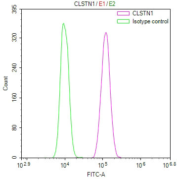
Species Reactivity
Human, Mouse, Rat
Immunogen
A synthesized peptide from human CLSTN1 protein
Immunogen Species
Homo sapiens (Human)
Conjugate
Non-conjugated
Clonality
Monoclonal
Isotype
Rabbit IgG
Clone No.
11H4
Purification Method
Affinity-chromatography
Concentration
It differs from different batches. Please contact us to confirm it.
Buffer
Rabbit IgG in 10mM phosphate buffered saline , pH 7.4, 150mM sodium chloride, 0.05% BSA, 0.02% sodium azide and 50% glycerol.
Form
Liquid
Tested Applications
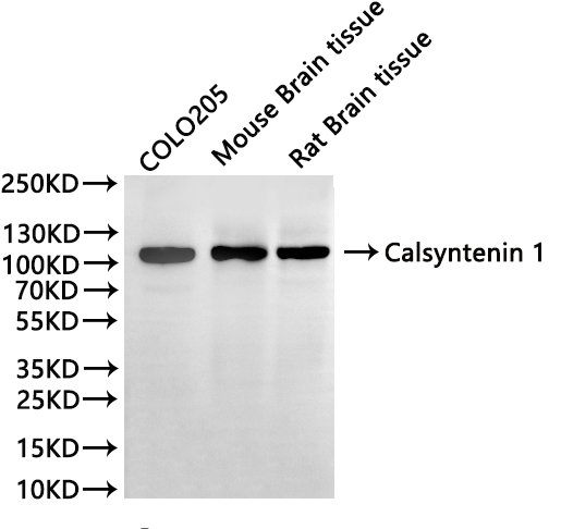
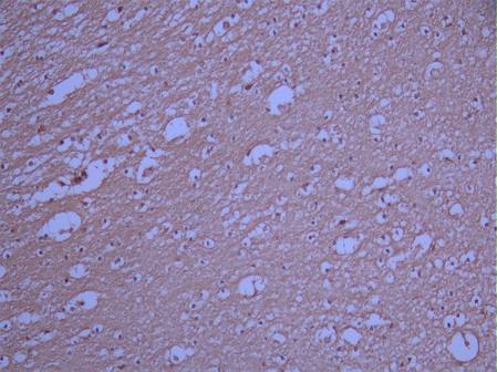
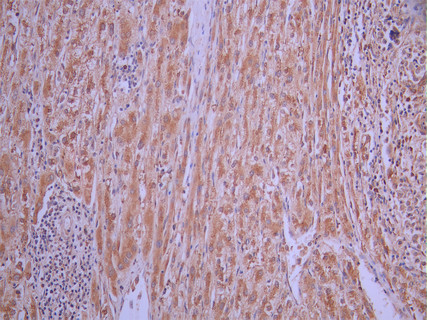
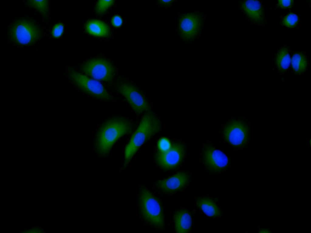
ELISA, WB, IHC, IF, FC
Recommended Dilution
Application Recommended Dilution
WB 1:500-1:5000
IHC 1:50-1:200
IF 1:50-1:200
FC 1:50-1:200
Troubleshooting and FAQs
Storage
Upon receipt, store at -20°C or -80°C. Avoid repeated freeze.
Lead Time
Basically, we can dispatch the products out in 1-3 working days after receiving your orders. Delivery time maybe differs from different purchasing way or location, please kindly consult your local distributors for specific delivery time.
Usage
For Research Use Only. Not for use in diagnostic or therapeutic procedures.

Customer Reviews and Q&A

 Customer Reviews

There are currently no reviews for this product.

Submit a Review here

Target Background

Function
Induces KLC1 association with vesicles and functions as a cargo in axonal anterograde transport. Complex formation with APBA2 and APP, stabilizes APP metabolism and enhances APBA2-mediated suppression of beta-APP40 secretion, due to the retardation of intracellular APP maturation. In complex with APBA2 and C99, a C-terminal APP fragment, abolishes C99 interaction with PSEN1 and thus APP C99 cleavage by gamma-secretase, most probably through stabilization of the direct interaction between APBA2 and APP. The intracellular fragment AlcICD suppresses APBB1-dependent transactivation stimulated by APP C-terminal intracellular fragment (AICD), most probably by competing with AICD for APBB1-binding. May modulate calcium-mediated postsynaptic signals.
Gene References into Functions
  1. Results suggests that APP partially compensates for defective Alcalpha in anterograde transport by providing an alternative cargo receptor for kinesin-1. PMID: 29093024
  2. Calsyntenin-1 contributes to the early stages of the hepatitis C virus replication cycle and the formation of the replication complex. PMID: 27221318
  3. Increased plasma p3-Alcalpha35, a major fragment of alcalpha1, may indicate an endophenotype in Alzheimer's disease patients with progressive cognitive impairment. PMID: 24305499
  4. Increased plasma p3-alcadein (alc) alpha, evident in the early stages of cognitive impairment, suggests that alc metabolites are useful plasma biomarkers of Alzheimer's disease. PMID: 22571980
  5. findings show that calsyntenin-1 is reduced in Alzheimer's disease brains and that the extent of this reduction correlates with increased amyloid-beta PMID: 22434822
  6. CLSTN1 is a target gene of the BACH1 transcription factor according to ChIP-seq analysis in HEK 293 cells. PMID: 21555518
  7. Phosphorylation of KLC1 at serine 460 modulates binding and trafficking of calsyntenin-1. PMID: 21385839
  8. Alcadein and amyloid beta-protein precursor regulates FE65-dependent gene transactivation [alcalpha1, alcbeta, alcgamma] PMID: 15037614

Show More

Hide All

Subcellular Location
Endoplasmic reticulum membrane; Single-pass type I membrane protein. Golgi apparatus membrane. Cell projection, neuron projection. Cell junction, synapse, postsynaptic cell membrane; Single-pass type I membrane protein. Nucleus.
Tissue Specificity
Expressed in the brain and, a lower level, in the heart, skeletal muscle, kidney and placenta. Accumulates in dystrophic neurites around the amyloid core of Alzheimer disease senile plaques (at protein level).
Database Links

HGNC: 17447

OMIM: 611321

KEGG: hsa:22883

STRING: 9606.ENSP00000366513

UniGene: Hs.29665

icon of phone
Call us
301-363-4651 (Available 9 a.m. to 5 p.m. CST from Monday to Friday)
icon of address
Address
7505 Fannin St., Ste 610, Room 7 (CUBIO Innovation Center), Houston, TX 77054, USA
icon of social media
Join us with

Subscribe newsletter

Leave a message

* To protect against spam, please pass the CAPTCHA test below.
CAPTCHA verification
© 2007-2025 CUSABIO TECHNOLOGY LLC All rights reserved. 鄂ICP备15011166号-1
×
Place an order now

I. Product details

*
*
*
*

II. Contact details

*
*

III. Ship To

*
*
*
*
*
*
*

IV. Bill To

*
*
*
*
*
*
*
*