EXOC3 Recombinant Monoclonal Antibody

Code CSB-RA585966A0HU
Size US$210
Order now
Image
  • IHC image of CSB-RA585966A0HU diluted at 1:100 and staining in paraffin-embedded human cervical cancer performed on a Leica BondTM system. After dewaxing and hydration, antigen retrieval was mediated by high pressure in a citrate buffer (pH 6.0). Section was blocked with 10% normal goat serum 30min at RT. Then primary antibody (1% BSA) was incubated at 4°C overnight. The primary is detected by a Goat anti-rabbit polymer IgG labeled by HRP and visualized using 0.05% DAB.
  • IHC image of CSB-RA585966A0HU diluted at 1:100 and staining in paraffin-embedded human skeletal muscle tissue performed on a Leica BondTM system. After dewaxing and hydration, antigen retrieval was mediated by high pressure in a citrate buffer (pH 6.0). Section was blocked with 10% normal goat serum 30min at RT. Then primary antibody (1% BSA) was incubated at 4°C overnight. The primary is detected by a Goat anti-rabbit polymer IgG labeled by HRP and visualized using 0.05% DAB.
  • Immunofluorescence staining of HepG2 cell with CSB-RA585966A0HU at 1:50, counter-stained with DAPI. The cells were fixed in 4% formaldehyde, permeabilized using 0.2% Triton X-100 and blocked in 10% normal Goat Serum. The cells were then incubated with the antibody overnight at 4°C. The secondary antibody was Alexa Fluor 488-congugated AffiniPure Goat Anti-Rabbit IgG(H+L).
  • Overlay Peak curve showing Hela cells stained with CSB-RA585966A0HU (red line) at 1:100. The cells were fixed in 4% formaldehyde and permeated by 0.2% TritonX-100 for 10min. Then 10% normal goat serum to block non-specific protein-protein interactions followed by the antibody (1ug/1*106cells) for 45min at 4℃. The secondary antibody used was FITC-conjugated goat anti-rabbit IgG (H+L) at 1/200 dilution for 35min at 4℃.Control antibody (green line) was Rabbit IgG (1ug/1*106cells) used under the same conditions. Acquisition of >10,000 events was performed.
Have Questions? Leave a Message or Start an on-line Chat

Product Details

Uniprot No.
Target Names
EXOC3
Alternative Names
Exocyst complex component 3 (Exocyst complex component Sec6), EXOC3, SEC6 SEC6L1
Species Reactivity
Human
Immunogen
A synthesized peptide from human EXOC3 protein
Immunogen Species
Homo sapiens (Human)
Conjugate
Non-conjugated
Clonality
Monoclonal
Isotype
Rabbit IgG
Clone No.
11B2
Purification Method
Affinity-chromatography
Concentration
It differs from different batches. Please contact us to confirm it.
Buffer
Preservative: 0.03% Proclin 300
Constituents: 50% Glycerol, 0.01M PBS, PH 7.4
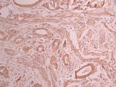
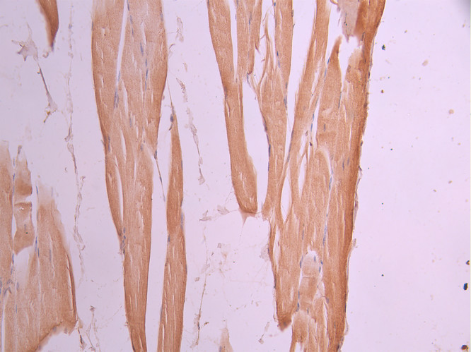
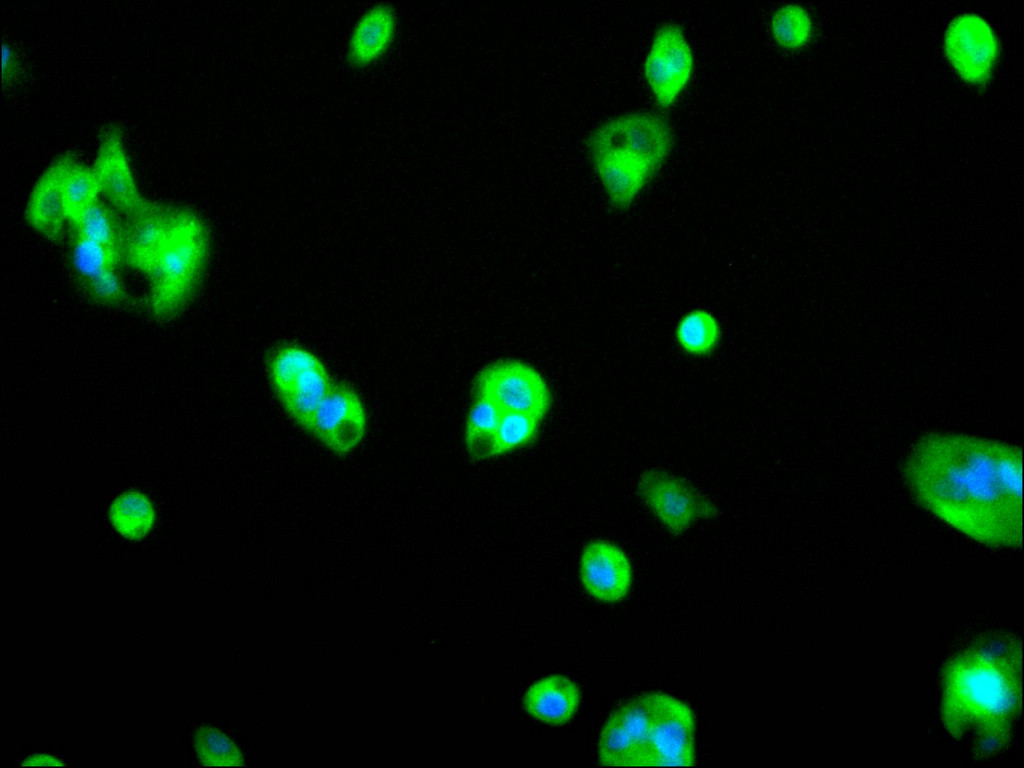
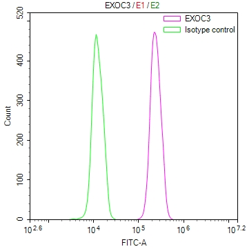
Form
Liquid
Tested Applications
ELISA, IHC, IF, FC
Recommended Dilution
Application Recommended Dilution
IHC 1:50-1:200
IF 1:50-1:200
FC 1:50-1:200
Troubleshooting and FAQs
Storage
Upon receipt, store at -20°C or -80°C. Avoid repeated freeze.
Lead Time
Basically, we can dispatch the products out in 1-3 working days after receiving your orders. Delivery time maybe differs from different purchasing way or location, please kindly consult your local distributors for specific delivery time.
Usage
For Research Use Only. Not for use in diagnostic or therapeutic procedures.

Customer Reviews and Q&A

 Customer Reviews

There are currently no reviews for this product.

Submit a Review here

Target Background

Function
Component of the exocyst complex involved in the docking of exocytic vesicles with fusion sites on the plasma membrane.
Gene References into Functions
  1. Sec6 regulate Bcl-2 and Mcl-1 expressions but not Bcl-xl in malignant peripheral nerve sheath tumor cells. PMID: 26892009
  2. Sec6 regulates NF-kappaB transcriptional activity via the control of the phosphorylation of IkappaBalpha, p90RSK1, and ERK PMID: 26247921
  3. The study explores the role of the exocyst complex component Sec6/8 in genomic stability. PMID: 26283729
  4. Sec6 regulates cytoplasmic translocation of p27 through p27 phosphorylation at Thr157, thereby promoting p27 degradation in the cytoplasm via interaction with Jab1 and Siah1 and suppressing cell cycle progression. PMID: 24949832
  5. Data demonstrate that the expression of alpha-E-catenin is increased by Sec6 siRNAs, and E-cadherin and beta-catenin localize mainly at the cell-cell contact region in HSC3 cells, which were transfected with Sec6 siRNA. PMID: 22381337

Show More

Hide All

Subcellular Location
Cytoplasm. Cytoplasm, perinuclear region. Cell projection, growth cone. Midbody. Golgi apparatus. Cell projection, neuron projection.
Protein Families
SEC6 family
Tissue Specificity
Expressed in epididymis (at protein level).
Database Links

HGNC: 30378

OMIM: 608186

KEGG: hsa:11336

STRING: 9606.ENSP00000323377

UniGene: Hs.481464

icon of phone
Call us
301-363-4651 (Available 9 a.m. to 5 p.m. CST from Monday to Friday)
icon of address
Address
7505 Fannin St., Ste 610, Room 7 (CUBIO Innovation Center), Houston, TX 77054, USA
icon of social media
Join us with

Subscribe newsletter

Leave a message

* To protect against spam, please pass the CAPTCHA test below.
CAPTCHA verification
© 2007-2026 CUSABIO TECHNOLOGY LLC All rights reserved. 鄂ICP备15011166号-1
Place an order now

I. Product details

*
*
*
*

II. Contact details

*
*

III. Ship To

*
*
*
*
*
*
*

IV. Bill To

*
*
*
*
*
*
*
*