FDX1 Recombinant Monoclonal Antibody

Code CSB-RA577140A0HU
Size US$210
Order now
Image
  • Western Blot
    Positive WB detected in: HEK293 whole cell lysate(30µg), A549 whole cell lysate(30µg), U-87MG whole cell lysate(30µg), Mouse kidney tissue lysate(30µg)
    All lanes: Ferredoxin 1 antibody at 1:1000
    Secondary
    Goat polyclonal to rabbit IgG at 1/40000 dilution
    Predicted band size: 19 kDa
    Observed band size: 13 kDa
    Exposure time:2min
  • IHC image of CSB-RA577140A0HU diluted at 1:100 and staining in paraffin-embedded human adrenal gland tissue performed on a Leica BondTM system. After dewaxing and hydration, antigen retrieval was mediated by high pressure in a citrate buffer (pH 6.0). Section was blocked with 10% normal goat serum 30min at RT. Then primary antibody (1% BSA) was incubated at 4°C overnight. The primary is detected by a Goat anti-rabbit polymer IgG labeled by HRP and visualized using 0.05% DAB.
  • IHC image of CSB-RA577140A0HU diluted at 1:100 and staining in paraffin-embedded human colorectal cancer performed on a Leica BondTM system. After dewaxing and hydration, antigen retrieval was mediated by high pressure in a citrate buffer (pH 6.0). Section was blocked with 10% normal goat serum 30min at RT. Then primary antibody (1% BSA) was incubated at 4°C overnight. The primary is detected by a Goat anti-rabbit polymer IgG labeled by HRP and visualized using 0.05% DAB.
  • Immunofluorescence staining of HepG2 cell with CSB-RA577140A0HU at 1:50 , counter-stained with DAPI. The cells were fixed in 4% formaldehyde, permeabilized using 0.2% Triton X-100 and blocked in 10% normal Goat Serum. The cells were then incubated with the antibody overnight at 4°C. The secondary antibody was Alexa Fluor 488-congugated AffiniPure Goat Anti-Rabbit IgG(H+L).
  • Overlay Peak curve showing Hela cells stained with CSB-RA577140A0HU (red line) at 1:100. The cells were fixed in 4% formaldehyde and permeated by 0.2% TritonX-100 for 10min. Then 10% normal goat serum to block non-specific protein-protein interactions followed by the antibody (1ug/1*106cells) for 45min at 4℃. The secondary antibody used was FITC-conjugated goat anti-rabbit IgG (H+L) at 1/200 dilution for 35min at 4℃.Control antibody (green line) was Rabbit IgG (1ug/1*106cells) used under the same conditions. Acquisition of >10, 000 events was performed.
Have Questions? Leave a Message or Start an on-line Chat

Product Details

Uniprot No.
Target Names
FDX1
Alternative Names
Adrenal ferredoxin; Adrenodoxin; Adrenodoxin; mitochondrial; ADX; ADX_HUMAN; FDX; fdx1; Ferredoxin 1; Ferredoxin-1; Hepatoredoxin; LOH11CR1D; Mitochondrial adrenodoxin; mitochondrial
Species Reactivity
Human, Mouse
Immunogen
A synthesized peptide from human FDX1 protein
Immunogen Species
Homo sapiens (Human)
Conjugate
Non-conjugated
Clonality
Monoclonal
Isotype
Rabbit IgG
Clone No.
8D3
Purification Method
Affinity-chromatography
Concentration
It differs from different batches. Please contact us to confirm it.
Buffer
Rabbit IgG in 10mM phosphate buffered saline , pH 7.4, 150mM sodium chloride, 0.05% BSA, 0.02% sodium azide and 50% glycerol.
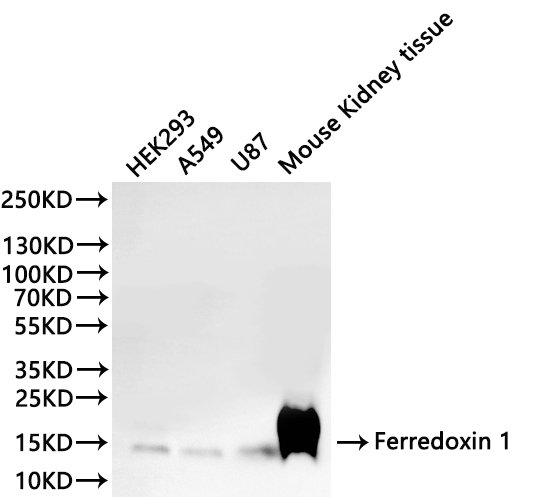
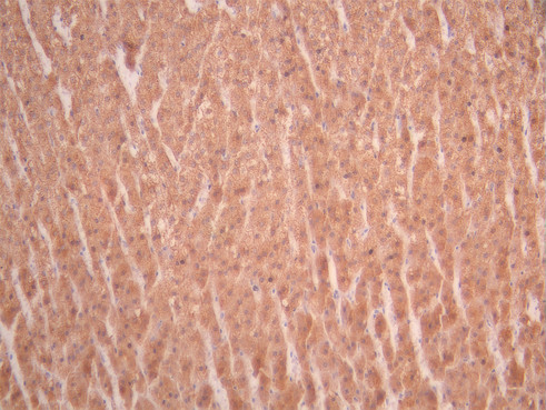
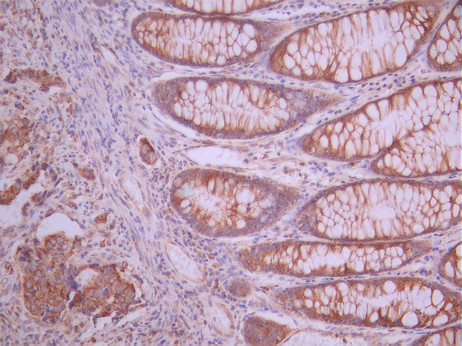
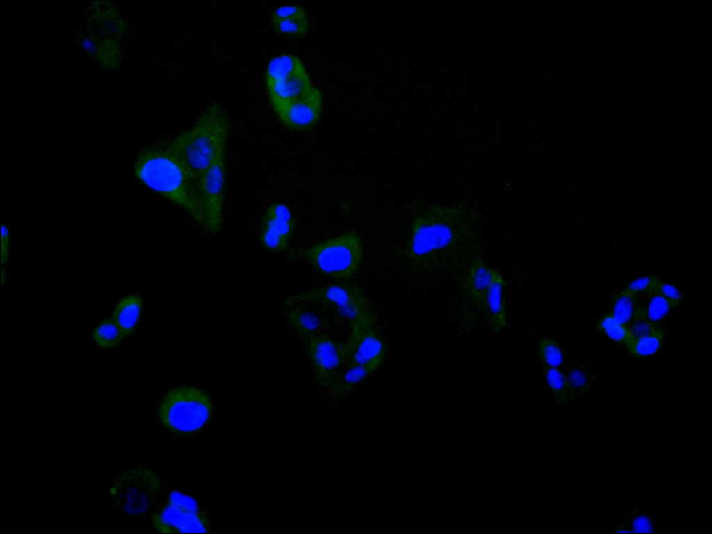
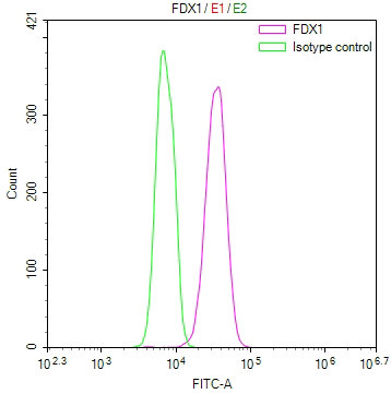
Form
Liquid
Tested Applications
ELISA, WB, IHC, IF, FC
Recommended Dilution
Application Recommended Dilution
WB 1:500-1:5000
IHC 1:50-1:200
IF 1:50-1:200
FC 1:50-1:200
Troubleshooting and FAQs
Storage
Upon receipt, store at -20°C or -80°C. Avoid repeated freeze.
Lead Time
Basically, we can dispatch the products out in 1-3 working days after receiving your orders. Delivery time maybe differs from different purchasing way or location, please kindly consult your local distributors for specific delivery time.
Usage
For Research Use Only. Not for use in diagnostic or therapeutic procedures.

Customer Reviews and Q&A

 Customer Reviews

There are currently no reviews for this product.

Submit a Review here

Target Background

Function
Essential for the synthesis of various steroid hormones. Participates in the reduction of mitochondrial cytochrome P450 for steroidogenesis. Transfers electrons from adrenodoxin reductase to CYP11A1, a cytochrome P450 that catalyzes cholesterol side-chain cleavage. Does not form a ternary complex with adrenodoxin reductase and CYP11A1 but shuttles between the two enzymes to transfer electrons.
Gene References into Functions
  1. Active Site Structures of CYP11A1 in the Presence of Its Physiological Substrates and Alterations upon Binding of Adrenodoxin PMID: 28991453
  2. Data suggest that binding sites between CYP11B1/CYP11B2 and adrenodoxin/ferredoxin-1 exhibit electrostatic interactions at K370 in CYP11B1 and at K366 in CYP11B2 mutant R366K with D79 in adrenodoxin/ferredoxin-1. (CYP11B1 = cytochrome P450 family 11 subfamily B member 1; CYP11B2 = cytochrome P450 family 11 subfamily B member 2) PMID: 28355486
  3. FDX1 and FDX2 both bind NFS1 and donate electrons for iron-sulfur cluster biosynthesis. PMID: 28001042
  4. TNFSF13 and FDX1 have potential roles in IgAN in the Han Chinese population. This information may be useful in the development of early prognostics for IgAN. PMID: 26431901
  5. Did not find any positive association between FDX1 SNPs and the risk of IgA nephropathy after adjustment for age and sex, but did find a significant and strong correlation with relevant clinical pathological parameters. PMID: 26370181
  6. results indicate transcription of FDX1 is regulated by the NR5A family and cAMP signaling, and participates in steroid hormone production in ovarian granulosa ce PMID: 23435367
  7. Data suggest that interference with any of the three related genes, ferredoxin (FDX)1, FDX2 or FDXR, disrupts iron-sulfur cluster assembly and maintenance of normal cytosolic and mitochondrial iron homeostasis. PMID: 22101253
  8. Results present the crystal structure of the complex of human adrenodoxin and CYP11A1--the first of a complex between a eukaryotic CYP and its redox partner. PMID: 21636783
  9. Unlike Fdx1, Fdx2 was unable to efficiently reduce mitochondrial cytochromes P450 and convert steroids, indicating that the two ferredoxin isoforms are highly specific for their substrates in distinct biochemical pathways. PMID: 20547883
  10. ADX rate of hydroxylation was linear with incubation time. PMID: 12782149
  11. Adrenodoxin induces apoptosis by the generation of reactive oxygen species in mitochondria PMID: 15927889
  12. Adx is able to support reactions catalyzed by human microsomal P450s: full length CYP17, truncated CYP17, and truncated CYP21 PMID: 17188650

Show More

Hide All

Subcellular Location
Mitochondrion matrix.
Protein Families
Adrenodoxin/putidaredoxin family
Tissue Specificity
Highest levels in the adrenal gland (at protein level). Also detected in kidney and testis (at protein level).
Database Links

HGNC: 3638

OMIM: 103260

KEGG: hsa:2230

STRING: 9606.ENSP00000260270

UniGene: Hs.744

icon of phone
Call us
301-363-4651 (Available 9 a.m. to 5 p.m. CST from Monday to Friday)
icon of address
Address
7505 Fannin St., Ste 610, Room 7 (CUBIO Innovation Center), Houston, TX 77054, USA
icon of social media
Join us with

Subscribe newsletter

Leave a message

* To protect against spam, please pass the CAPTCHA test below.
CAPTCHA verification
© 2007-2025 CUSABIO TECHNOLOGY LLC All rights reserved. 鄂ICP备15011166号-1
×
Place an order now

I. Product details

*
*
*
*

II. Contact details

*
*

III. Ship To

*
*
*
*
*
*
*

IV. Bill To

*
*
*
*
*
*
*
*